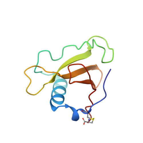
2SAR

DETERMINATION AND RESTRAINED LEAST-SQUARES REFINEMENT OF THE CRYSTAL STRUCTURES OF RIBONUCLEASE SA AND ITS COMPLEX WITH 3'-GUANYLIC ACID AT 1.8 ANGSTROMS RESOLUTION


Experimental Data Snapshot

  • Method: X-RAY DIFFRACTION
  • Resolution: 1.80 Å
  • R-Value Observed: 0.175 

wwPDB Validation   3D Report Full Report


Ligand Structure Quality Assessment 


This is version 1.4 of the entry. See complete history


Literature

Determination and restrained least-squares refinement of the structures of ribonuclease Sa and its complex with 3'-guanylic acid at 1.8 A resolution.

Sevcik, J.Dodson, E.J.Dodson, G.G.

(1991) Acta Crystallogr B 47: 240-253

  • Primary Citation of Related Structures:  
    1SAR, 2SAR

  • PubMed Abstract: 

    The crystal structures of ribonuclease from Streptomyces aureofaciens (RNase Sa) and its complex with 3'-guanylic acid (guanosine 3'-monophosphate, 3'-GMP) have been determined by the method of isomorphous replacement. The atomic parameters have been refined by restrained least-squares minimization using data in the resolution range 10.0-1.8 A. All protein atoms and more than 230 water atoms in the two crystal structures have been refined to crystallographic R factors of 0.172 and 0.175 respectively. The estimated r.m.s. error in the atomic positions ranges from 0.2 A for well-defined atoms to about 0.5 A for more poorly defined atoms. There are two enzyme molecules in the asymmetric unit, built independently, and referred to as molecules A and B. The value of the average B factor for protein atoms in both structures is about 19 A2 and for water molecules about 35 A2. Electron density for the substrate analogue 3'-GMP was found only at the active site of molecule A. The density was very clear and the positions of all 3'-GMP atoms were refined with precision comparable to that of the protein.


  • Organizational Affiliation

    Institute of Molecular Biology, Slovak Academy of Sciences, Bratislava, Czechoslovakia.


Macromolecules
Find similar proteins by:  (by identity cutoff)  |  3D Structure
Entity ID: 1
MoleculeChains Sequence LengthOrganismDetailsImage
RIBONUCLEASE SA
A, B
96Kitasatospora aureofaciensMutation(s): 0 
EC: 4.6.1.24
UniProt
Find proteins for P05798 (Kitasatospora aureofaciens)
Explore P05798 
Go to UniProtKB:  P05798
Entity Groups  
Sequence Clusters30% Identity50% Identity70% Identity90% Identity95% Identity100% Identity
UniProt GroupP05798
Sequence Annotations
Expand
  • Reference Sequence
Experimental Data & Validation

Experimental Data

  • Method: X-RAY DIFFRACTION
  • Resolution: 1.80 Å
  • R-Value Observed: 0.175 
  • Space Group: P 21 21 21
Unit Cell:
Length ( Å )Angle ( ˚ )
a = 64.9α = 90
b = 78.32β = 90
c = 38.79γ = 90
Software Package:
Software NamePurpose
PROLSQrefinement

Structure Validation

View Full Validation Report


Ligand Structure Quality Assessment 


Entry History 

Deposition Data

Revision History  (Full details and data files)

  • Version 1.0: 1992-04-15
    Type: Initial release
  • Version 1.1: 2008-03-03
    Changes: Version format compliance
  • Version 1.2: 2011-07-13
    Changes: Version format compliance
  • Version 1.3: 2017-11-29
    Changes: Derived calculations, Other
  • Version 1.4: 2024-10-09
    Changes: Data collection, Database references, Derived calculations, Structure summary