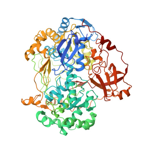
2VPZ

Polysulfide reductase native structure


Experimental Data Snapshot

  • Method: X-RAY DIFFRACTION
  • Resolution: 2.40 Å
  • R-Value Free: 0.253 
  • R-Value Work: 0.249 
  • R-Value Observed: 0.249 

wwPDB Validation   3D Report Full Report


Ligand Structure Quality Assessment 


This is version 1.3 of the entry. See complete history


Literature

Molecular Mechanism of Energy Conservation in Polysulfide Respiration

Jormakka, M.Yokoyama, K.Yano, T.Tamakoshi, M.Akimoto, S.Shimamura, T.Curmi, P.Iwata, S.

(2008) Nat Struct Mol Biol 15: 730

  • DOI: https://doi.org/10.1038/nsmb.1434
  • Primary Citation of Related Structures:  
    2VPW, 2VPX, 2VPY, 2VPZ

  • PubMed Abstract: 

    Bacterial polysulfide reductase (PsrABC) is an integral membrane protein complex responsible for quinone-coupled reduction of polysulfide, a process important in extreme environments such as deep-sea vents and hot springs. We determined the structure of polysulfide reductase from Thermus thermophilus at 2.4-A resolution, revealing how the PsrA subunit recognizes and reduces its unique polyanionic substrate. The integral membrane subunit PsrC was characterized using the natural substrate menaquinone-7 and inhibitors, providing a comprehensive representation of a quinone binding site and revealing the presence of a water-filled cavity connecting the quinone binding site on the periplasmic side to the cytoplasm. These results suggest that polysulfide reductase could be a key energy-conserving enzyme of the T. thermophilus respiratory chain, using polysulfide as the terminal electron acceptor and pumping protons across the membrane via a previously unknown mechanism.


  • Organizational Affiliation

    Department of Biophysics, University of New South Wales, Barker Street, Sydney, New South Wales 2052, Australia. [email protected]


Macromolecules
Find similar proteins by:  (by identity cutoff)  |  3D Structure
Entity ID: 1
MoleculeChains Sequence LengthOrganismDetailsImage
THIOSULFATE REDUCTASEA,
D [auth E]
765Thermus thermophilus HB27Mutation(s): 0 
EC: 1
UniProt
Find proteins for Q72LA4 (Thermus thermophilus (strain ATCC BAA-163 / DSM 7039 / HB27))
Explore Q72LA4 
Go to UniProtKB:  Q72LA4
Entity Groups  
Sequence Clusters30% Identity50% Identity70% Identity90% Identity95% Identity100% Identity
UniProt GroupQ72LA4
Sequence Annotations
Expand
  • Reference Sequence
Find similar proteins by:  (by identity cutoff)  |  3D Structure
Entity ID: 2
MoleculeChains Sequence LengthOrganismDetailsImage
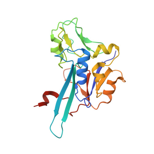
NRFC PROTEINB,
E [auth F]
195Thermus thermophilus HB27Mutation(s): 0 
UniProt
Find proteins for Q72LA5 (Thermus thermophilus (strain ATCC BAA-163 / DSM 7039 / HB27))
Explore Q72LA5 
Go to UniProtKB:  Q72LA5
Entity Groups  
Sequence Clusters30% Identity50% Identity70% Identity90% Identity95% Identity100% Identity
UniProt GroupQ72LA5
Sequence Annotations
Expand
  • Reference Sequence
Find similar proteins by:  (by identity cutoff)  |  3D Structure
Entity ID: 3
MoleculeChains Sequence LengthOrganismDetailsImage
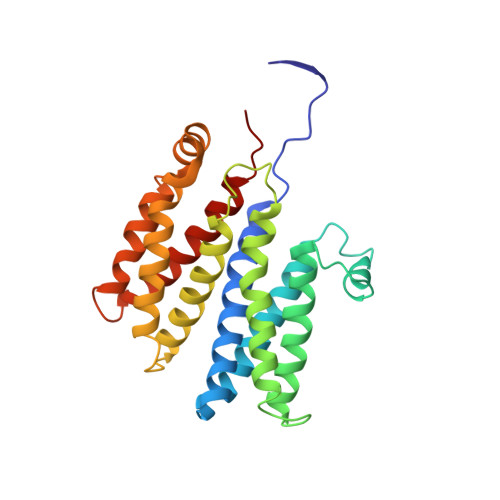
HYPOTHETICAL MEMBRANE SPANNING PROTEINC,
F [auth G]
253Thermus thermophilus HB27Mutation(s): 0 
Membrane Entity: Yes 
UniProt
Find proteins for Q72LA6 (Thermus thermophilus (strain ATCC BAA-163 / DSM 7039 / HB27))
Explore Q72LA6 
Go to UniProtKB:  Q72LA6
Entity Groups  
Sequence Clusters30% Identity50% Identity70% Identity90% Identity95% Identity100% Identity
UniProt GroupQ72LA6
Sequence Annotations
Expand
  • Reference Sequence
Small Molecules
Ligands 3 Unique
IDChains Name / Formula / InChI Key2D Diagram3D Interactions
MGD
Query on MGD

Download Ideal Coordinates CCD File 
H [auth A],
I [auth A],
P [auth E],
Q [auth E]
2-AMINO-5,6-DIMERCAPTO-7-METHYL-3,7,8A,9-TETRAHYDRO-8-OXA-1,3,9,10-TETRAAZA-ANTHRACEN-4-ONE GUANOSINE DINUCLEOTIDE
C20 H26 N10 O13 P2 S2
VQAGYJCYOLHZDH-ILXWUORBSA-N
SF4
Query on SF4

Download Ideal Coordinates CCD File 
G [auth A]
K [auth B]
L [auth B]
M [auth B]
N [auth B]
G [auth A],
K [auth B],
L [auth B],
M [auth B],
N [auth B],
O [auth E],
S [auth F],
T [auth F],
U [auth F],
V [auth F]
IRON/SULFUR CLUSTER
Fe4 S4
LJBDFODJNLIPKO-UHFFFAOYSA-N
MO
Query on MO

Download Ideal Coordinates CCD File 
J [auth A],
R [auth E]
MOLYBDENUM ATOM
Mo
ZOKXTWBITQBERF-UHFFFAOYSA-N
Experimental Data & Validation

Experimental Data

  • Method: X-RAY DIFFRACTION
  • Resolution: 2.40 Å
  • R-Value Free: 0.253 
  • R-Value Work: 0.249 
  • R-Value Observed: 0.249 
  • Space Group: P 21 21 21
Unit Cell:
Length ( Å )Angle ( ˚ )
a = 118.405α = 90
b = 166.265β = 90
c = 246.428γ = 90
Software Package:
Software NamePurpose
CNSrefinement
MOSFLMdata reduction
SCALAdata scaling
REFMACphasing

Structure Validation

View Full Validation Report


Ligand Structure Quality Assessment 


Entry History 

Deposition Data

Revision History  (Full details and data files)

  • Version 1.0: 2008-06-10
    Type: Initial release
  • Version 1.1: 2011-05-08
    Changes: Version format compliance
  • Version 1.2: 2011-07-13
    Changes: Version format compliance
  • Version 1.3: 2024-05-08
    Changes: Data collection, Database references, Derived calculations, Other