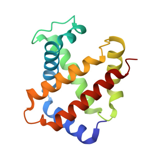
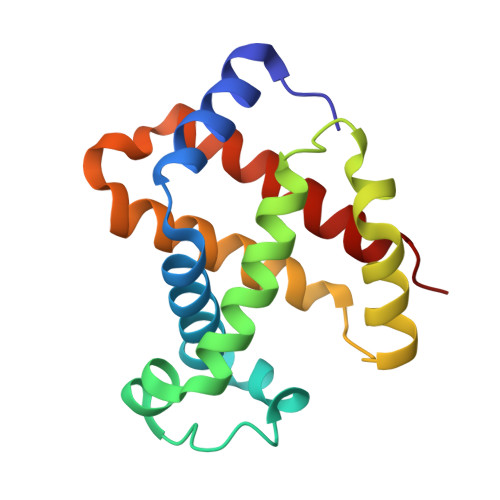
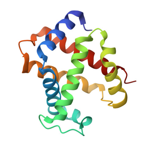
2W72

DEOXYGENATED STRUCTURE OF A DISTAL SITE HEMOGLOBIN MUTANT PLUS XE


Experimental Data Snapshot

  • Method: X-RAY DIFFRACTION
  • Resolution: 1.07 Å
  • R-Value Free: 0.153 
  • R-Value Work: 0.129 
  • R-Value Observed: 0.130 

Starting Model: experimental
View more details

wwPDB Validation   3D Report Full Report


Ligand Structure Quality Assessment 


This is version 1.4 of the entry. See complete history


Literature

Pattern of Cavities in Globins: The Case of Human Hemoglobin.

Savino, C.Miele, A.E.Draghi, F.Johnson, K.A.Sciara, G.Brunori, M.Vallone, B.

(2009) Biopolymers 91: 1097

  • DOI: https://doi.org/10.1002/bip.21201
  • Primary Citation of Related Structures:  
    2W6V, 2W6W, 2W72

  • PubMed Abstract: 

    Our aim is to shed light on the conservation of potential ligand docking sites that play an important role in ligand dynamics of globins by using the technique of filling internal cavities naturally present in hemoglobin and myoglobin with xenon atoms. In particular, we present the high resolution structures of the Xe-adduct of deoxygenated wild type human hemoglobin and a quadruple mutant (L(B10)Y and H(E7)Q in alpha and beta chains). For the sake of comparison we also determined under the same experimental conditions the xenon complex of wild type sperm whale myoglobin. The analysis revealed that the number and position of Xe binding cavities are different in the alpha and beta subunits, the latter being more similar to myoglobin. Notably, no proximal Xe docking site was detected in hemoglobin, at variance with myoglobin. The pattern of internal cavities accessibility and affinity for xenon suggests a different role for the dynamics of ligand migration in the two types of hemoglobin chains as compared to myoglobin. The number and position of hydrophobic cavities in hemoglobin are briefly discussed also in comparison with the data available for other members of the globin superfamily.


  • Organizational Affiliation

    University of Rome, P.le Aldo Moro 5, 00185 Rome, Italy. [email protected]


Macromolecules
Find similar proteins by:  (by identity cutoff)  |  3D Structure
Entity ID: 1
MoleculeChains Sequence LengthOrganismDetailsImage
HUMAN HEMOGLOBIN A141Homo sapiensMutation(s): 2 
UniProt & NIH Common Fund Data Resources
Find proteins for P69905 (Homo sapiens)
Explore P69905 
Go to UniProtKB:  P69905
PHAROS:  P69905
Entity Groups  
Sequence Clusters30% Identity50% Identity70% Identity90% Identity95% Identity100% Identity
UniProt GroupP69905
Sequence Annotations
Expand
  • Reference Sequence
Find similar proteins by:  (by identity cutoff)  |  3D Structure
Entity ID: 2
MoleculeChains Sequence LengthOrganismDetailsImage
HUMAN HEMOGLOBIN A
B, D
146Homo sapiensMutation(s): 2 
UniProt & NIH Common Fund Data Resources
Find proteins for P68871 (Homo sapiens)
Explore P68871 
Go to UniProtKB:  P68871
PHAROS:  P68871
GTEx:  ENSG00000244734 
Entity Groups  
Sequence Clusters30% Identity50% Identity70% Identity90% Identity95% Identity100% Identity
UniProt GroupP68871
Sequence Annotations
Expand
  • Reference Sequence
Find similar proteins by:  (by identity cutoff)  |  3D Structure
Entity ID: 3
MoleculeChains Sequence LengthOrganismDetailsImage
HUMAN HEMOGLOBIN A141Homo sapiensMutation(s): 2 
UniProt & NIH Common Fund Data Resources
Find proteins for P69905 (Homo sapiens)
Explore P69905 
Go to UniProtKB:  P69905
PHAROS:  P69905
Entity Groups  
Sequence Clusters30% Identity50% Identity70% Identity90% Identity95% Identity100% Identity
UniProt GroupP69905
Sequence Annotations
Expand
  • Reference Sequence
Small Molecules
Ligands 5 Unique
IDChains Name / Formula / InChI Key2D Diagram3D Interactions
HEM
Query on HEM

Download Ideal Coordinates CCD File 
E [auth A],
FA [auth D],
P [auth B],
V [auth C]
PROTOPORPHYRIN IX CONTAINING FE
C34 H32 Fe N4 O4
KABFMIBPWCXCRK-RGGAHWMASA-L
XE
Query on XE

Download Ideal Coordinates CCD File 
AA [auth C]
BA [auth C]
CA [auth C]
DA [auth C]
EA [auth C]
AA [auth C],
BA [auth C],
CA [auth C],
DA [auth C],
EA [auth C],
HA [auth D],
I [auth A],
IA [auth D],
J [auth A],
K [auth A],
L [auth A],
M [auth A],
N [auth A],
T [auth B],
U [auth B]
XENON
Xe
FHNFHKCVQCLJFQ-UHFFFAOYSA-N
SO4
Query on SO4

Download Ideal Coordinates CCD File 
F [auth A]
G [auth A]
GA [auth D]
H [auth A]
Q [auth B]
F [auth A],
G [auth A],
GA [auth D],
H [auth A],
Q [auth B],
R [auth B],
W [auth C],
X [auth C]
SULFATE ION
O4 S
QAOWNCQODCNURD-UHFFFAOYSA-L
PO4
Query on PO4

Download Ideal Coordinates CCD File 
Y [auth C]PHOSPHATE ION
O4 P
NBIIXXVUZAFLBC-UHFFFAOYSA-K
K
Query on K

Download Ideal Coordinates CCD File 
O [auth A],
S [auth B],
Z [auth C]
POTASSIUM ION
K
NPYPAHLBTDXSSS-UHFFFAOYSA-N
Experimental Data & Validation

Experimental Data

  • Method: X-RAY DIFFRACTION
  • Resolution: 1.07 Å
  • R-Value Free: 0.153 
  • R-Value Work: 0.129 
  • R-Value Observed: 0.130 
  • Space Group: P 1 21 1
Unit Cell:
Length ( Å )Angle ( ˚ )
a = 61.957α = 90
b = 82.447β = 99.6
c = 53.531γ = 90
Software Package:
Software NamePurpose
REFMACrefinement
DENZOdata reduction
SCALAdata scaling
MOLREPphasing

Structure Validation

View Full Validation Report


Ligand Structure Quality Assessment 


Entry History 

Deposition Data

Revision History  (Full details and data files)

  • Version 1.0: 2009-04-28
    Type: Initial release
  • Version 1.1: 2011-08-17
    Changes: Database references, Derived calculations, Non-polymer description, Other, Version format compliance
  • Version 1.2: 2019-07-24
    Changes: Data collection, Derived calculations
  • Version 1.3: 2023-03-08
    Changes: Database references, Derived calculations, Other
  • Version 1.4: 2024-02-07
    Changes: Data collection, Refinement description