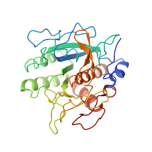
2WUV

Crystallographic analysis of counter-ion effects on subtilisin enzymatic action in acetonitrile


Experimental Data Snapshot

  • Method: X-RAY DIFFRACTION
  • Resolution: 2.24 Å
  • R-Value Free: 0.231 
  • R-Value Work: 0.160 
  • R-Value Observed: 0.163 

Starting Model: experimental
View more details

wwPDB Validation   3D Report Full Report


This is version 1.3 of the entry. See complete history


Literature

Crystallographic Analysis of Counterion Effects on Subtilisin Enzymatic Action in Acetonitrile.

Cianci, M.Tomaszewski, B.Helliwell, J.R.Halling, P.J.

(2010) J Am Chem Soc 132: 2293

  • DOI: https://doi.org/10.1021/ja908703c
  • Primary Citation of Related Structures:  
    2WUV, 2WUW

  • PubMed Abstract: 

    When enzymes are in low dielectric nonaqueous media, it would be expected that their charged groups would be more closely associated with counterions. There is evidence that these counterions may then affect enzymatic activity. Published crystal structures of proteins in organic solvents do not show increased numbers of associated counterions, and this might reflect the difficulty of distinguishing cations like Na(+) from water molecules. In this paper, the placement of several Cs(+) and Cl(-) ions in crystals of the serine protease subtilisin Carlsberg is presented. Ions are more readily identified crystallographically through their anomalous diffraction using softer X-rays. The protein conformation is very similar to that of the enzyme without CsCl in acetonitrile, both for the previously reported ( 1SCB ) and our own newly determined model. No fewer than 11 defined sites for Cs(+) cations and 8 Cl(-) anions are identified around the protein molecule, although most of these have partial occupancy and may represent nonspecific binding sites. Two Cs(+) and two Cl(-) ions are close to the mouth of the active site cleft, where they may affect catalysis. In fact, cross-linked CsCl-treated subtilisin crystals transferred to acetonitrile show catalytic activity several fold higher than the reference crystals containing Na(+). Presoaking with another large cation, choline, also increases the enzyme activity. The active site appears only minimally sterically perturbed by the ion presence around it, so alternative activation mechanisms can be suggested: an electrostatic redistribution and/or a larger hydration sphere that enhances the protein domain.


  • Organizational Affiliation

    European Molecular Biology Laboratory, Hamburg Outstation, c/o DESY, Building 25a, Notkestrasse 85, 22603 Hamburg, Germany. [email protected]


Macromolecules
Find similar proteins by:  (by identity cutoff)  |  3D Structure
Entity ID: 1
MoleculeChains Sequence LengthOrganismDetailsImage
SUBTILISIN CARLSBERG274Bacillus licheniformisMutation(s): 0 
EC: 3.4.21.62
UniProt
Find proteins for P00780 (Bacillus licheniformis)
Explore P00780 
Go to UniProtKB:  P00780
Entity Groups  
Sequence Clusters30% Identity50% Identity70% Identity90% Identity95% Identity100% Identity
UniProt GroupP00780
Sequence Annotations
Expand
  • Reference Sequence
Small Molecules
Ligands 5 Unique
IDChains Name / Formula / InChI Key2D Diagram3D Interactions
CS
Query on CS

Download Ideal Coordinates CCD File 
I [auth A]
J [auth A]
K [auth A]
L [auth A]
M [auth A]
I [auth A],
J [auth A],
K [auth A],
L [auth A],
M [auth A],
N [auth A],
O [auth A],
P [auth A],
Q [auth A],
R [auth A],
S [auth A]
CESIUM ION
Cs
NCMHKCKGHRPLCM-UHFFFAOYSA-N
CCN
Query on CCN

Download Ideal Coordinates CCD File 
V [auth A],
W [auth A],
X [auth A],
Y [auth A],
Z [auth A]
ACETONITRILE
C2 H3 N
WEVYAHXRMPXWCK-UHFFFAOYSA-N
CA
Query on CA

Download Ideal Coordinates CCD File 
T [auth A]CALCIUM ION
Ca
BHPQYMZQTOCNFJ-UHFFFAOYSA-N
CL
Query on CL

Download Ideal Coordinates CCD File 
AA [auth A]
B [auth A]
C [auth A]
D [auth A]
E [auth A]
AA [auth A],
B [auth A],
C [auth A],
D [auth A],
E [auth A],
F [auth A],
G [auth A],
H [auth A]
CHLORIDE ION
Cl
VEXZGXHMUGYJMC-UHFFFAOYSA-M
NA
Query on NA

Download Ideal Coordinates CCD File 
U [auth A]SODIUM ION
Na
FKNQFGJONOIPTF-UHFFFAOYSA-N
Experimental Data & Validation

Experimental Data

  • Method: X-RAY DIFFRACTION
  • Resolution: 2.24 Å
  • R-Value Free: 0.231 
  • R-Value Work: 0.160 
  • R-Value Observed: 0.163 
  • Space Group: P 21 21 21
Unit Cell:
Length ( Å )Angle ( ˚ )
a = 52.177α = 90
b = 55.311β = 90
c = 75.778γ = 90
Software Package:
Software NamePurpose
REFMACrefinement
HKL-2000data reduction
HKL-2000data scaling
MOLREPphasing

Structure Validation

View Full Validation Report


Entry History 

Deposition Data

Revision History  (Full details and data files)

  • Version 1.0: 2010-12-08
    Type: Initial release
  • Version 1.1: 2011-05-08
    Changes: Version format compliance
  • Version 1.2: 2011-07-13
    Changes: Version format compliance
  • Version 1.3: 2023-12-20
    Changes: Data collection, Database references, Derived calculations, Other, Refinement description