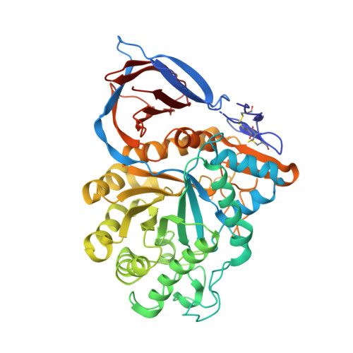
2XWD

X-RAY STRUCTURE OF ACID-BETA-GLUCOSIDASE WITH 5N,6O-(N'-(N-OCTYL)IMINO)NOJIRIMYCIN IN THE ACTIVE SITE


Experimental Data Snapshot

  • Method: X-RAY DIFFRACTION
  • Resolution: 2.66 Å
  • R-Value Free: 0.219 
  • R-Value Work: 0.150 
  • R-Value Observed: 0.153 

Starting Model: experimental
View more details

wwPDB Validation   3D Report Full Report


Ligand Structure Quality Assessment 


This is version 2.2 of the entry. See complete history


Literature

Cyclodextrin-Mediated Crystallization of Acid Beta-Glucosidase in Complex with Amphiphilic Bicyclic Nojirimycin Analogues.

Brumshtein, B.Aguilar-Moncayo, M.Benito, J.M.Garcia Fernandez, J.M.Silman, I.Shaaltiel, Y.Aviezer, D.Sussman, J.L.Futerman, A.H.Ortiz Mellet, C.

(2011) Org Biomol Chem 9: 4160

  • DOI: https://doi.org/10.1039/c1ob05200d
  • Primary Citation of Related Structures:  
    2XWD, 2XWE

  • PubMed Abstract: 

    Cyclodextrin-based host-guest chemistry has been exploited to facilitate co-crystallization of recombinant human acid β-glucosidase (β-glucocerebrosidase, GlcCerase) with amphiphilic bicyclic nojirimycin analogues of the sp(2)-iminosugar type. Attempts to co-crystallize GlcCerase with 5-N,6-O-[N'-(n-octyl)iminomethylidene]nojirimycin (NOI-NJ) or with 5-N,6-S-[N'-(n-octyl)iminomethylidene]-6-thionojirimycin (6S-NOI-NJ), two potent inhibitors of the enzyme with promising pharmacological chaperone activity for several Gaucher disease-associated mutations, were unsuccessful probably due to the formation of aggregates that increase the heterogeneity of the sample and affect nucleation and growth of crystals. Cyclomaltoheptaose (β-cyclodextrin, βCD) efficiently captures NOI-NJ and 6S-NOI-NJ in aqueous media to form inclusion complexes in which the lipophilic tail is accommodated in the hydrophobic cavity of the cyclooligosaccharide. The dissociation constant of the complex of the amphiphilic sp(2)-iminosugars with βCD is two orders of magnitude higher than that of the corresponding complex with GlcCerase, allowing the efficient transfer of the inhibitor from the βCD cavity to the GlcCerase active site. Enzyme-inhibitor complexes suitable for X-ray analysis were thus grown in the presence of βCD. In contrast to what was previously observed for the complex of GlcCerase with the more basic derivative, 6-amino-6-deoxy-5-N,6-N-[N'-(n-octyl)iminomethylidene]nojirimycin (6N-NOI-NJ), the β-anomers of both NOI-NJ and 6S-NOI-NJ were seen in the active site, even though the α-anomer was exclusively detected both in aqueous solution and in the corresponding βCD:sp(2)-iminosugar complexes. Our results further suggest that cyclodextrin derivatives might serve as suitable delivery systems of amphiphilic glycosidase inhibitors in a biomedical context.


  • Organizational Affiliation

    Department of Structural Biology, Weizmann Institute of Science, Rehovot, 76100, Israel.


Macromolecules
Find similar proteins by:  (by identity cutoff)  |  3D Structure
Entity ID: 1
MoleculeChains Sequence LengthOrganismDetailsImage
GLUCOSYLCERAMIDASE
A, B
505Homo sapiensMutation(s): 1 
EC: 3.2.1.45 (PDB Primary Data), 3.2.1.46 (UniProt), 2.4.1 (UniProt), 3.2.1 (UniProt)
UniProt & NIH Common Fund Data Resources
Find proteins for P04062 (Homo sapiens)
Explore P04062 
Go to UniProtKB:  P04062
PHAROS:  P04062
GTEx:  ENSG00000177628 
Entity Groups  
Sequence Clusters30% Identity50% Identity70% Identity90% Identity95% Identity100% Identity
UniProt GroupP04062
Glycosylation
Glycosylation Sites: 1Go to GlyGen: P04062-1
Sequence Annotations
Expand
  • Reference Sequence
Oligosaccharides

Help

Entity ID: 2
MoleculeChains Length2D Diagram Glycosylation3D Interactions
alpha-D-mannopyranose-(1-4)-2-acetamido-2-deoxy-beta-D-glucopyranose-(1-4)-[alpha-L-fucopyranose-(1-3)]2-acetamido-2-deoxy-beta-D-glucopyranose
C
4N-Glycosylation
Glycosylation Resources
GlyTouCan:  G73622RM
GlyCosmos:  G73622RM
GlyGen:  G73622RM
Entity ID: 3
MoleculeChains Length2D Diagram Glycosylation3D Interactions
beta-D-mannopyranose-(1-4)-2-acetamido-2-deoxy-beta-D-glucopyranose-(1-4)-[alpha-L-fucopyranose-(1-3)]2-acetamido-2-deoxy-beta-D-glucopyranose
D
4N-Glycosylation
Glycosylation Resources
GlyTouCan:  G18638YB
GlyCosmos:  G18638YB
GlyGen:  G18638YB
Small Molecules
Ligands 2 Unique
IDChains Name / Formula / InChI Key2D Diagram3D Interactions
LGS
Query on LGS

Download Ideal Coordinates CCD File 
J [auth A],
P [auth B]
(3Z,5S,6R,7S,8R,8aR)-3-(octylimino)hexahydro[1,3]oxazolo[3,4-a]pyridine-5,6,7,8-tetrol
C15 H28 N2 O5
QJILQIWQVOAQBB-KRIYVDMXSA-N
SO4
Query on SO4

Download Ideal Coordinates CCD File 
E [auth A]
F [auth A]
G [auth A]
H [auth A]
I [auth A]
E [auth A],
F [auth A],
G [auth A],
H [auth A],
I [auth A],
K [auth B],
L [auth B],
M [auth B],
N [auth B],
O [auth B]
SULFATE ION
O4 S
QAOWNCQODCNURD-UHFFFAOYSA-L
Binding Affinity Annotations 
IDSourceBinding Affinity
LGS PDBBind:  2XWD Ki: 110 (nM) from 1 assay(s)
Experimental Data & Validation

Experimental Data

  • Method: X-RAY DIFFRACTION
  • Resolution: 2.66 Å
  • R-Value Free: 0.219 
  • R-Value Work: 0.150 
  • R-Value Observed: 0.153 
  • Space Group: P 1 21 1
Unit Cell:
Length ( Å )Angle ( ˚ )
a = 68.036α = 90
b = 96.617β = 103.69
c = 83.164γ = 90
Software Package:
Software NamePurpose
REFMACrefinement
HKL-2000data reduction
SCALEPACKdata scaling
PHASERphasing

Structure Validation

View Full Validation Report


Ligand Structure Quality Assessment 


Entry History 

Revision History  (Full details and data files)

  • Version 1.0: 2011-09-14
    Type: Initial release
  • Version 2.0: 2020-07-29
    Type: Remediation
    Reason: Carbohydrate remediation
    Changes: Advisory, Atomic model, Data collection, Derived calculations, Other, Structure summary
  • Version 2.1: 2023-12-20
    Changes: Data collection, Database references, Derived calculations, Refinement description, Structure summary
  • Version 2.2: 2024-10-09
    Changes: Structure summary