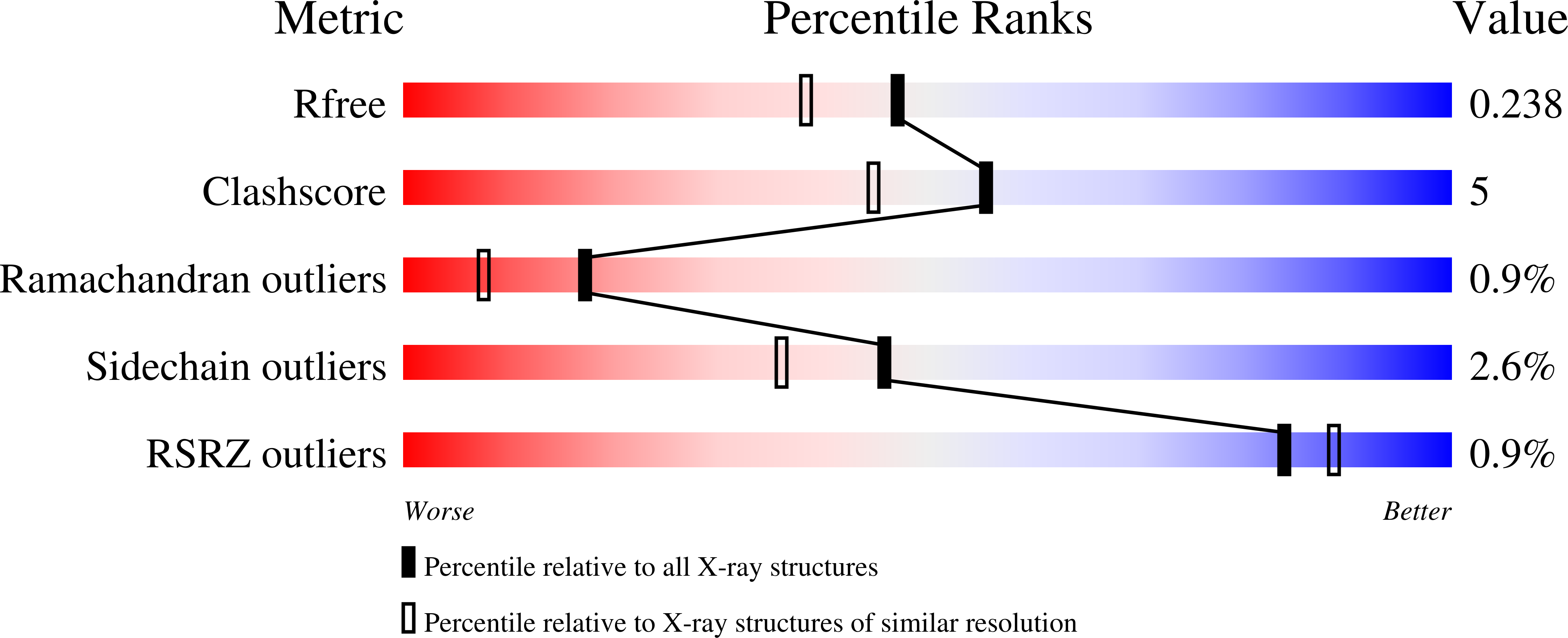
2Z49

Crystal Structure of Hemolytic Lectin CEL-III Complexed with methyl-alpha-D-galactopylanoside


Experimental Data Snapshot

  • Method: X-RAY DIFFRACTION
  • Resolution: 1.95 Å
  • R-Value Free: 0.239 
  • R-Value Work: 0.191 
  • R-Value Observed: 0.193 

Starting Model: experimental
View more details

wwPDB Validation   3D Report Full Report


Ligand Structure Quality Assessment 


This is version 2.3 of the entry. See complete history


Literature

C-type lectin-like carbohydrate-recognition of the hemolytic lectin CEL-III containing ricin-type beta-trefoil folds

Hatakeyama, T.Unno, H.Kouzuma, Y.Uchida, T.Eto, S.Hidemura, H.Kato, N.Yonekura, M.Kusunoki, M.

(2007) J Biol Chem 282: 37826-37835

  • DOI: https://doi.org/10.1074/jbc.M705604200
  • Primary Citation of Related Structures:  
    2Z48, 2Z49

  • PubMed Abstract: 

    CEL-III is a Ca(2+)-dependent hemolytic lectin, isolated from the marine invertebrate Cucumaria echinata. The three-dimensional structure of CEL-III/GalNAc and CEL-III/methyl alpha-galactoside complexes was solved by x-ray crystallographic analysis. In these complexes, five carbohydrate molecules were found to be bound to two carbohydrate-binding domains (domains 1 and 2) located in the N-terminal 2/3 portion of the polypeptide and that contained beta-trefoil folds similar to ricin B-chain. The 3-OH and 4-OH of bound carbohydrate molecules were coordinated with Ca(2+) located at the subdomains 1alpha, 1gamma, 2alpha, 2beta, and 2gamma, simultaneously forming hydrogen bond networks with nearby amino acid side chains, which is similar to carbohydrate binding in C-type lectins. The binding of carbohydrates was further stabilized by aromatic amino acid residues, such as tyrosine and tryptophan, through a stacking interaction with the hydrophobic face of carbohydrates. The importance of amino acid residues in the carbohydrate-binding sites was confirmed by the mutational analyses. The orientation of bound GalNAc and methyl alpha-galactoside was similar to the galactose moiety of lactose bound to the carbohydrate-binding site of the ricin B-chain, although the ricin B-chain does not require Ca(2+) ions for carbohydrate binding. The binding of the carbohydrates induced local structural changes in carbohydrate-binding sites in subdomains 2alpha and 2beta. Binding of GalNAc also induced a slight change in the main chain structure of domain 3, which could be related to the conformational change upon binding of specific carbohydrates to induce oligomerization of the protein.


  • Organizational Affiliation

    Department of Applied Chemistry, Faculty of Engineering, Nagasaki University, Nagasaki, Japan. [email protected]


Macromolecules
Find similar proteins by:  (by identity cutoff)  |  3D Structure
Entity ID: 1
MoleculeChains Sequence LengthOrganismDetailsImage
Hemolytic lectin CEL-III
A, B
432Cucumaria echinataMutation(s): 0 
UniProt
Find proteins for Q868M7 (Cucumaria echinata)
Explore Q868M7 
Go to UniProtKB:  Q868M7
Entity Groups  
Sequence Clusters30% Identity50% Identity70% Identity90% Identity95% Identity100% Identity
UniProt GroupQ868M7
Sequence Annotations
Expand
  • Reference Sequence
Small Molecules
Ligands 3 Unique
IDChains Name / Formula / InChI Key2D Diagram3D Interactions
AMG
Query on AMG

Download Ideal Coordinates CCD File 
C [auth A]
D [auth A]
E [auth A]
F [auth A]
G [auth A]
C [auth A],
D [auth A],
E [auth A],
F [auth A],
G [auth A],
O [auth B],
P [auth B],
Q [auth B],
R [auth B],
S [auth B]
methyl alpha-D-galactopyranoside
C7 H14 O6
HOVAGTYPODGVJG-PZRMXXKTSA-N
CA
Query on CA

Download Ideal Coordinates CCD File 
H [auth A]
I [auth A]
J [auth A]
K [auth A]
L [auth A]
H [auth A],
I [auth A],
J [auth A],
K [auth A],
L [auth A],
T [auth B],
U [auth B],
V [auth B],
W [auth B],
X [auth B]
CALCIUM ION
Ca
BHPQYMZQTOCNFJ-UHFFFAOYSA-N
MG
Query on MG

Download Ideal Coordinates CCD File 
M [auth A],
N [auth A],
Y [auth B],
Z [auth B]
MAGNESIUM ION
Mg
JLVVSXFLKOJNIY-UHFFFAOYSA-N
Modified Residues  1 Unique
IDChains TypeFormula2D DiagramParent
PCA
Query on PCA
A, B
L-PEPTIDE LINKINGC5 H7 N O3GLN
Experimental Data & Validation

Experimental Data

  • Method: X-RAY DIFFRACTION
  • Resolution: 1.95 Å
  • R-Value Free: 0.239 
  • R-Value Work: 0.191 
  • R-Value Observed: 0.193 
  • Space Group: P 1 21 1
Unit Cell:
Length ( Å )Angle ( ˚ )
a = 53.287α = 90
b = 65.451β = 97.07
c = 126.993γ = 90
Software Package:
Software NamePurpose
REFMACrefinement
MOSFLMdata reduction
SCALAdata scaling
PHASERphasing

Structure Validation

View Full Validation Report


Ligand Structure Quality Assessment 


Entry History 

Deposition Data

Revision History  (Full details and data files)

  • Version 1.0: 2007-10-30
    Type: Initial release
  • Version 1.1: 2011-07-13
    Changes: Version format compliance
  • Version 2.0: 2019-12-25
    Changes: Derived calculations, Polymer sequence
  • Version 2.1: 2020-07-29
    Type: Remediation
    Reason: Carbohydrate remediation
    Changes: Data collection, Derived calculations, Structure summary
  • Version 2.2: 2023-11-01
    Changes: Data collection, Database references, Refinement description, Structure summary
  • Version 2.3: 2024-10-23
    Changes: Structure summary