2ZMA

Crystal Structure of 6-Aminohexanoate-dimer Hydrolase S112A/G181D/H266N/D370Y Mutant with Substrate


Experimental Data Snapshot

  • Method: X-RAY DIFFRACTION
  • Resolution: 1.51 Å
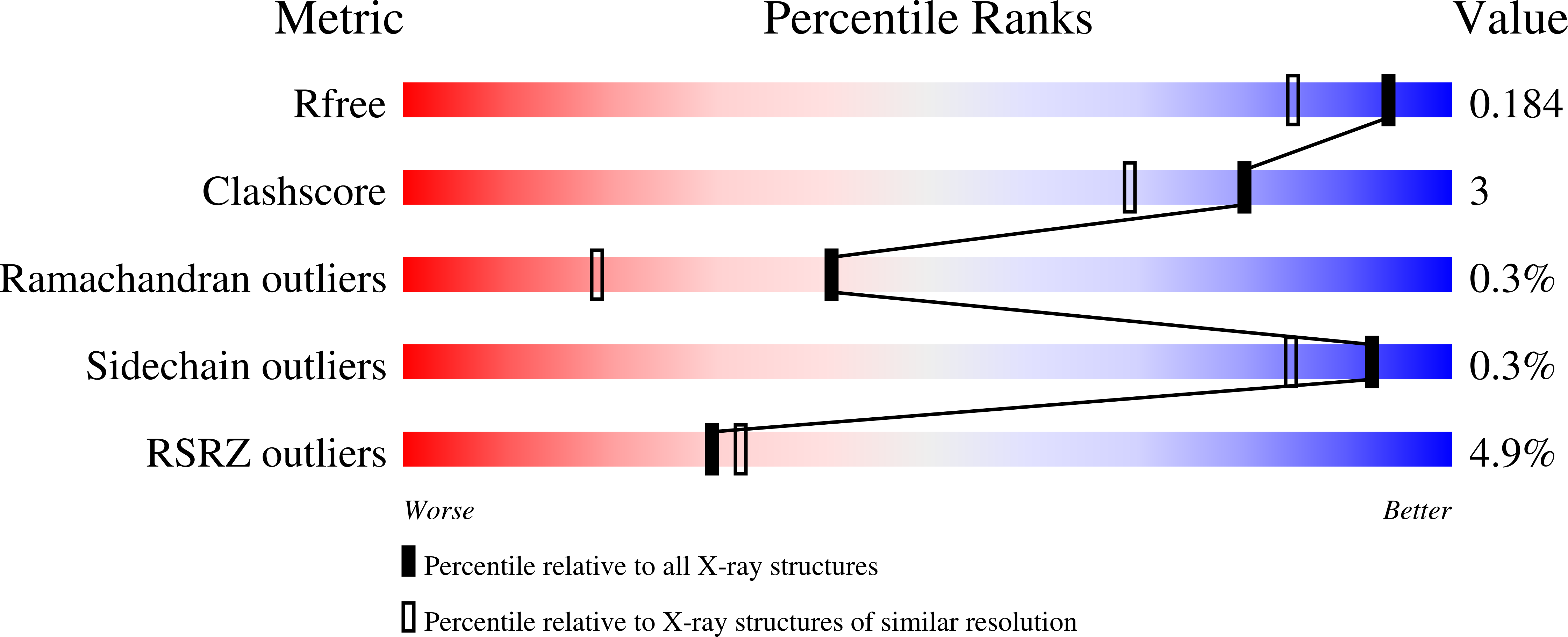
  • R-Value Free: 0.190 
  • R-Value Work: 0.175 
  • R-Value Observed: 0.175 

Starting Model: experimental
View more details

wwPDB Validation   3D Report Full Report


This is version 2.0 of the entry. See complete history


Literature

Molecular design of a nylon-6 byproduct-degrading enzyme from a carboxylesterase with a beta-lactamase fold

Kawashima, Y.Ohki, T.Shibata, N.Higuchi, Y.Wakitani, Y.Matsuura, Y.Nakata, Y.Takeo, M.Kato, D.Negoro, S.

(2009) FEBS J 276: 2547-2556

  • DOI: https://doi.org/10.1111/j.1742-4658.2009.06978.x
  • Primary Citation of Related Structures:  
    2E8I, 2ZM0, 2ZM7, 2ZMA

  • PubMed Abstract: 

    A carboxylesterase with a beta-lactamase fold from Arthrobacter possesses a low level of hydrolytic activity (0.023 mumol.min(-1).mg(-1)) when acting on a 6-aminohexanoate linear dimer byproduct of the nylon-6 industry (Ald). G181D/H266N/D370Y triple mutations in the parental esterase increased the Ald-hydrolytic activity 160-fold. Kinetic studies showed that the triple mutant possesses higher affinity for the substrate Ald (K(m) = 2.0 mm) than the wild-type Ald hydrolase from Arthrobacter (K(m) = 21 mm). In addition, the k(cat)/K(m) of the mutant (1.58 s(-1).mm(-1)) was superior to that of the wild-type enzyme (0.43 s(-1).mm(-1)), demonstrating that the mutant efficiently converts the unnatural amide compounds even at low substrate concentrations, and potentially possesses an advantage for biotechnological applications. X-ray crystallographic analyses of the G181D/H266N/D370Y enzyme and the inactive S112A-mutant-Ald complex revealed that Ald binding induces rotation of Tyr370/His375, movement of the loop region (N167-V177), and flip-flop of Tyr170, resulting in the transition from open to closed forms. From the comparison of the three-dimensional structures of various mutant enzymes and site-directed mutagenesis at positions 266 and 370, we now conclude that Asn266 makes suitable contacts with Ald and improves the electrostatic environment at the N-terminal region of Ald cooperatively with Asp181, and that Tyr370 stabilizes Ald binding by hydrogen-bonding/hydrophobic interactions at the C-terminal region of Ald.


  • Organizational Affiliation

    Department of Materials Science and Chemistry, Graduate School of Engineering, University of Hyogo, Japan.


Macromolecules
Find similar proteins by:  (by identity cutoff)  |  3D Structure
Entity ID: 1
MoleculeChains Sequence LengthOrganismDetailsImage
6-aminohexanoate-dimer hydrolase392Flavobacterium sp.Mutation(s): 4 
Gene Names: nylBnylB'
EC: 3.5.1.46
UniProt
Find proteins for P07062 (Paenarthrobacter ureafaciens)
Explore P07062 
Go to UniProtKB:  P07062
Find proteins for P07061 (Paenarthrobacter ureafaciens)
Explore P07061 
Go to UniProtKB:  P07061
Entity Groups  
Sequence Clusters30% Identity50% Identity70% Identity90% Identity95% Identity100% Identity
UniProt GroupsP07061P07062
Sequence Annotations
Expand
  • Reference Sequence
Small Molecules
Ligands 4 Unique
IDChains Name / Formula / InChI Key2D Diagram3D Interactions
MES
Query on MES

Download Ideal Coordinates CCD File 
I [auth A]2-(N-MORPHOLINO)-ETHANESULFONIC ACID
C6 H13 N O4 S
SXGZJKUKBWWHRA-UHFFFAOYSA-N
ACA
Query on ACA

Download Ideal Coordinates CCD File 
G [auth A],
H [auth A]
6-AMINOHEXANOIC ACID
C6 H13 N O2
SLXKOJJOQWFEFD-UHFFFAOYSA-N
SO4
Query on SO4

Download Ideal Coordinates CCD File 
B [auth A],
C [auth A],
D [auth A],
E [auth A],
F [auth A]
SULFATE ION
O4 S
QAOWNCQODCNURD-UHFFFAOYSA-L
GOL
Query on GOL

Download Ideal Coordinates CCD File 
J [auth A],
K [auth A],
L [auth A],
M [auth A],
N [auth A]
GLYCEROL
C3 H8 O3
PEDCQBHIVMGVHV-UHFFFAOYSA-N
Experimental Data & Validation

Experimental Data

  • Method: X-RAY DIFFRACTION
  • Resolution: 1.51 Å
  • R-Value Free: 0.190 
  • R-Value Work: 0.175 
  • R-Value Observed: 0.175 
  • Space Group: P 32 2 1
Unit Cell:
Length ( Å )Angle ( ˚ )
a = 96.687α = 90
b = 96.687β = 90
c = 112.911γ = 120
Software Package:
Software NamePurpose
CNSrefinement
HKL-2000data reduction
SCALEPACKdata scaling
CNSphasing

Structure Validation

View Full Validation Report


Entry History 

Deposition Data

Revision History  (Full details and data files)

  • Version 1.0: 2009-04-07
    Type: Initial release
  • Version 1.1: 2011-07-13
    Changes: Non-polymer description, Version format compliance
  • Version 1.2: 2017-08-09
    Changes: Refinement description, Source and taxonomy
  • Version 1.3: 2021-11-10
    Changes: Database references, Derived calculations
  • Version 1.4: 2023-11-01
    Changes: Data collection, Refinement description
  • Version 2.0: 2023-11-15
    Changes: Atomic model, Data collection, Derived calculations