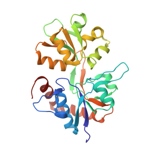
Binding and Selectivity of the Marine Toxin Neodysiherbaine A and Its Synthetic Analogues to GluK1 and GluK2 Kainate Receptors.
Unno, M., Shinohara, M., Takayama, K., Tanaka, H., Teruya, K., Doh-Ura, K., Sakai, R., Sasaki, M., Ikeda-Saito, M.(2011) J Mol Biol 413: 667-683
- PubMed: 21893069
- DOI: https://doi.org/10.1016/j.jmb.2011.08.043
- Primary Citation of Related Structures:
2ZNS, 2ZNT, 2ZNU, 3FUZ, 3FV1, 3FV2, 3FVG, 3FVK, 3FVN, 3QXM - PubMed Abstract:
Dysiherbaine (DH) and neodysiherbaine A (NDH) selectively bind and activate two kainate-type ionotropic glutamate receptors, GluK1 and GluK2. The ligand-binding domains of human GluK1 and GluK2 were crystallized as bound forms with a series of DH analogues including DH, NDH, 8-deoxy-NDH, 9-deoxy-NDH and 8,9-dideoxy-NDH (MSVIII-19), isolated from natural sources or prepared by total synthesis. Since the DH analogues exhibit a wide range of binding affinities and agonist efficacies, it follows that the detailed analysis of crystal structure would provide us with a significant opportunity to elucidate structural factors responsible for selective binding and some aspects of gating efficacy. We found that differences in three amino acids (Thr503, Ser706 and Ser726 in GluK1 and Ala487, Asn690 and Thr710 in GluK2) in the ligand-binding pocket generate differences in the binding modes of NDH to GluK1 and GluK2. Furthermore, deletion of the C(9) hydroxy group in NDH alters the ligand conformation such that it is no longer suited for binding to the GluK1 ligand-binding pocket. In GluK2, NDH pushes and rotates the side chain of Asn690 (substituted for Ser706 in GluK1) and disrupts an interdomain hydrogen bond with Glu409. The present data support the idea that receptor selectivities of DH analogues resulted from the differences in the binding modes of the ligands in GluK1/GluK2 and the steric repulsion of Asn690 in GluK2. All ligands, regardless of agonist efficacy, induced full domain closure. Consequently, ligand efficacy and domain closure did not directly coincide with DH analogues and the kainate receptors.
Organizational Affiliation:
Frontier Research Center for Applied Atomic Sciences, Ibaraki University, Tokai, Naka, Ibaraki 319-1106, Japan. [email protected]


