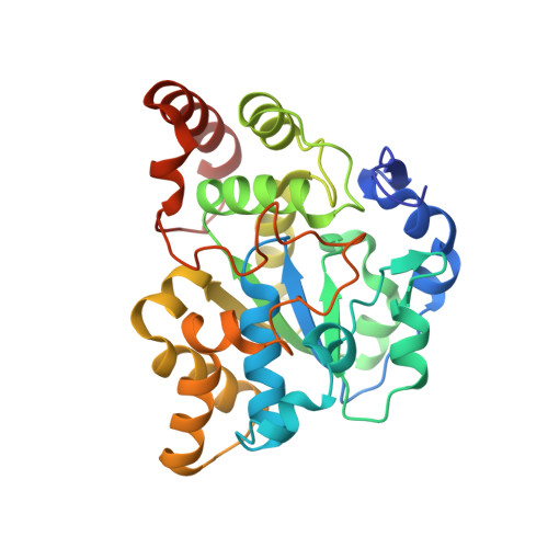
2ZPT

Crystal structure of mouse sulfotransferase SULT1D1 complex with PAP


Experimental Data Snapshot

  • Method: X-RAY DIFFRACTION
  • Resolution: 1.15 Å
  • R-Value Free: 0.160 
  • R-Value Work: 0.148 
  • R-Value Observed: 0.149 

Starting Model: experimental
View more details

wwPDB Validation   3D Report Full Report


Ligand Structure Quality Assessment 


This is version 1.2 of the entry. See complete history


Literature

Crystal structure of mSULT1D1, a mouse catecholamine sulfotransferase

Teramoto, T.Sakakibara, Y.Inada, K.Kurogi, K.Liu, M.-C.Suiko, M.Kimura, M.Kakuta, Y.

(2008) FEBS Lett 582: 3909-3914

  • DOI: https://doi.org/10.1016/j.febslet.2008.10.035
  • Primary Citation of Related Structures:  
    2ZPT

  • PubMed Abstract: 

    In mammals, sulfonation as mediated by specific cytosolic sulfotransferases (SULTs) plays an important role in the homeostasis of dopamine and other catecholamines. To gain insight into the structural basis for dopamine recognition/binding, we determined the crystal structure of a mouse dopamine-sulfating SULT, mouse SULT1D1 (mSULT1D1). Data obtained indicated that mSULT1D1 comprises of a single alpha/beta domain with a five-stranded parallel beta-sheet. In contrast to the structure of the human SULT1A3 (hSULT1A3)-dopamine complex previously reported, molecular modeling and mutational analysis revealed that a water molecule plays a critical role in the recognition of the amine group of dopamine by mSULT1D1. These results imply differences in substrate binding between dopamine-sulfating SULTs from different species.


  • Organizational Affiliation

    Laboratory of Structural Biology, Department of Systems Life Sciences, Japan.


Macromolecules
Find similar proteins by:  (by identity cutoff)  |  3D Structure
Entity ID: 1
MoleculeChains Sequence LengthOrganismDetailsImage
Tyrosine-ester sulfotransferaseA [auth X]295Mus musculusMutation(s): 0 
Gene Names: Sult1d1
EC: 2.8.2.9 (PDB Primary Data), 2.8.2 (UniProt)
UniProt
Find proteins for Q3UZZ6 (Mus musculus)
Explore Q3UZZ6 
Go to UniProtKB:  Q3UZZ6
Entity Groups  
Sequence Clusters30% Identity50% Identity70% Identity90% Identity95% Identity100% Identity
UniProt GroupQ3UZZ6
Sequence Annotations
Expand
  • Reference Sequence
Experimental Data & Validation

Experimental Data

  • Method: X-RAY DIFFRACTION
  • Resolution: 1.15 Å
  • R-Value Free: 0.160 
  • R-Value Work: 0.148 
  • R-Value Observed: 0.149 
  • Space Group: C 1 2 1
Unit Cell:
Length ( Å )Angle ( ˚ )
a = 155.819α = 90
b = 67.6β = 105.63
c = 42.696γ = 90
Software Package:
Software NamePurpose
REFMACrefinement
BBSdata collection
HKL-2000data reduction
HKL-2000data scaling
MOLREPphasing

Structure Validation

View Full Validation Report


Ligand Structure Quality Assessment 


Entry History 

Deposition Data

Revision History  (Full details and data files)

  • Version 1.0: 2008-11-18
    Type: Initial release
  • Version 1.1: 2011-07-13
    Changes: Non-polymer description, Version format compliance
  • Version 1.2: 2023-11-01
    Changes: Data collection, Database references, Derived calculations, Refinement description