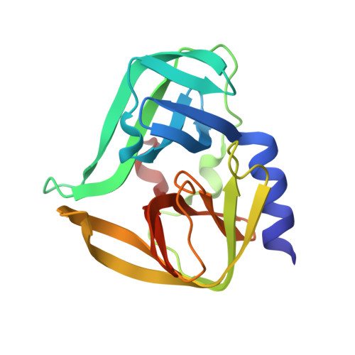
Structural Basis of Inhibition Specificities of 3C and 3C-like Proteases by Zinc-coordinating and Peptidomimetic Compounds
Lee, C.C., Kuo, C.J., Ko, T.P., Hsu, M.F., Tsui, Y.C., Chang, S.C., Yang, S., Chen, S.J., Chen, H.C., Hsu, M.C., Shih, S.R., Liang, P.H., Wang, A.H.-J.(2009) J Biol Chem 284: 7646-7655
- PubMed: 19144641
- DOI: https://doi.org/10.1074/jbc.M807947200
- Primary Citation of Related Structures:
2ZTX, 2ZTY, 2ZTZ, 2ZU1, 2ZU2, 2ZU3, 2ZU4, 2ZU5 - PubMed Abstract:
Human coxsackievirus (CV) belongs to the picornavirus family, which consists of over 200 medically relevant viruses. In picornavirus, a chymotrypsin-like protease (3C(pro)) is required for viral replication by processing the polyproteins, and thus it is regarded as an antiviral drug target. A 3C-like protease (3CL(pro)) also exists in human coronaviruses (CoV) such as 229E and the one causing severe acute respiratory syndrome (SARS). To combat SARS, we previously had developed peptidomimetic and zinc-coordinating inhibitors of 3CL(pro). As shown in the present study, some of these compounds were also found to be active against 3C(pro) of CV strain B3 (CVB3). Several crystal structures of 3C(pro) from CVB3 and 3CL(pro) from CoV-229E and SARS-CoV in complex with the inhibitors were solved. The zinc-coordinating inhibitor is tetrahedrally coordinated to the His(40)-Cys(147) catalytic dyad of CVB3 3C(pro). The presence of specific binding pockets for the residues of peptidomimetic inhibitors explains the binding specificity. Our results provide a structural basis for inhibitor optimization and development of potential drugs for antiviral therapies.
Organizational Affiliation:
Structural Biology Program, Institute of Biochemistry and Molecular Biology, National Yang-Ming University, Taipei 11221, Taiwan.


