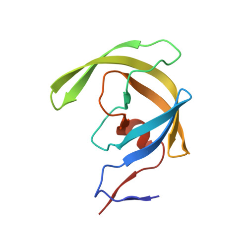
2ZYE

Structure of HIV-1 Protease in Complex with Potent Inhibitor KNI-272 Determined by Neutron Crystallography


Experimental Data Snapshot

  • Method: NEUTRON DIFFRACTION
  • Resolution: 1.90 Å
  • R-Value Free: 0.222 
  • R-Value Work: 0.193 
  • R-Value Observed: 0.194 

wwPDB Validation   3D Report Full Report


This is version 1.5 of the entry. See complete history


Literature

Structure of HIV-1 protease in complex with potent inhibitor KNI-272 determined by high-resolution X-ray and neutron crystallography.

Adachi, M.Ohhara, T.Kurihara, K.Tamada, T.Honjo, E.Okazaki, N.Arai, S.Shoyama, Y.Kimura, K.Matsumura, H.Sugiyama, S.Adachi, H.Takano, K.Mori, Y.Hidaka, K.Kimura, T.Hayashi, Y.Kiso, Y.Kuroki, R.

(2009) Proc Natl Acad Sci U S A 

  • DOI: https://doi.org/10.1073/pnas.0809400106
  • Primary Citation of Related Structures:  
    2ZYE, 3FX5

  • PubMed Abstract: 

    HIV-1 protease is a dimeric aspartic protease that plays an essential role in viral replication. To further understand the catalytic mechanism and inhibitor recognition of HIV-1 protease, we need to determine the locations of key hydrogen atoms in the catalytic aspartates Asp-25 and Asp-125. The structure of HIV-1 protease in complex with transition-state analog KNI-272 was determined by combined neutron crystallography at 1.9-A resolution and X-ray crystallography at 1.4-A resolution. The resulting structural data show that the catalytic residue Asp-25 is protonated and that Asp-125 (the catalytic residue from the corresponding diad-related molecule) is deprotonated. The proton on Asp-25 makes a hydrogen bond with the carbonyl group of the allophenylnorstatine (Apns) group in KNI-272. The deprotonated Asp-125 bonds to the hydroxyl proton of Apns. The results provide direct experimental evidence for proposed aspects of the catalytic mechanism of HIV-1 protease and can therefore contribute substantially to the development of specific inhibitors for therapeutic application.


  • Organizational Affiliation

    Molecular Structural Biology Group, Quantum Beam Science Directorate, Japan Atomic Energy Agency, 2-4 Shirakata-Shirane, Tokai, Ibaraki 319-1195, Japan.


Macromolecules
Find similar proteins by:  (by identity cutoff)  |  3D Structure
Entity ID: 1
MoleculeChains Sequence LengthOrganismDetailsImage
protease
A, B
99Human immunodeficiency virus 1Mutation(s): 5 
Gene Names: pol
EC: 3.4.23.16
UniProt
Find proteins for P03367 (Human immunodeficiency virus type 1 group M subtype B (isolate BRU/LAI))
Explore P03367 
Go to UniProtKB:  P03367
Entity Groups  
Sequence Clusters30% Identity50% Identity70% Identity90% Identity95% Identity100% Identity
UniProt GroupP03367
Sequence Annotations
Expand
  • Reference Sequence
Small Molecules
Ligands 1 Unique
IDChains Name / Formula / InChI Key2D Diagram3D Interactions
KNI
Query on KNI

Download Ideal Coordinates CCD File 
C [auth B](4R)-N-tert-butyl-3-[(2S,3S)-2-hydroxy-3-({N-[(isoquinolin-5-yloxy)acetyl]-S-methyl-L-cysteinyl}amino)-4-phenylbutanoyl]-1,3-thiazolidine-4-carboxamide
C33 H41 N5 O6 S2
NJBBLOIWMSYVCQ-VZTVMPNDSA-N
Binding Affinity Annotations 
IDSourceBinding Affinity
KNI BindingDB:  2ZYE Ki: 0.9 (nM) from 1 assay(s)
Biologically Interesting Molecules (External Reference) 1 Unique
Entity ID: 2
IDChains NameType/Class2D Diagram3D Interactions
PRD_000562 (KNI)
Query on PRD_000562
C [auth B]kynostatin 272Peptide-like / Enzyme inhibitor
Experimental Data & Validation

Experimental Data

  • Method: NEUTRON DIFFRACTION
  • Resolution: 1.90 Å
  • R-Value Free: 0.222 
  • R-Value Work: 0.193 
  • R-Value Observed: 0.194 
  • Space Group: P 21 21 2
Unit Cell:
Length ( Å )Angle ( ˚ )
a = 59.5α = 90
b = 87.4β = 90
c = 46.8γ = 90
Software Package:
Software NamePurpose
PHENIXrefinement
DENZOdata reduction
SCALEPACKdata scaling

Structure Validation

View Full Validation Report


Entry History 

Deposition Data

Revision History  (Full details and data files)

  • Version 1.0: 2009-03-24
    Type: Initial release
  • Version 1.1: 2011-07-13
    Changes: Atomic model, Database references, Derived calculations, Non-polymer description, Structure summary, Version format compliance
  • Version 1.2: 2012-12-12
    Changes: Other
  • Version 1.3: 2018-06-20
    Changes: Data collection
  • Version 1.4: 2021-11-10
    Changes: Database references, Derived calculations, Structure summary
  • Version 1.5: 2024-05-29
    Changes: Data collection