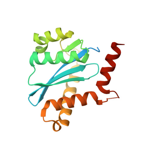
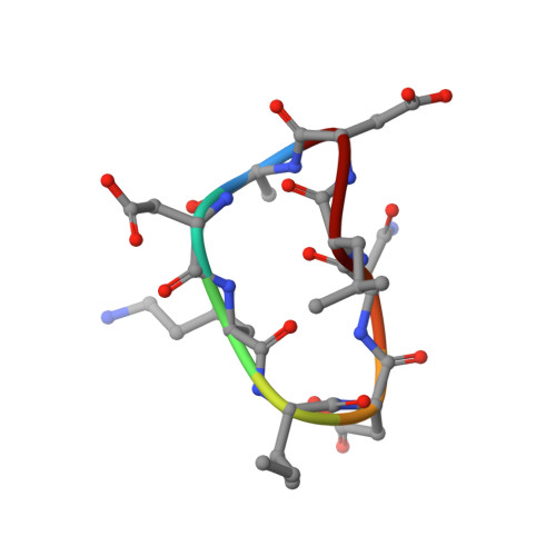
3AVG

Crystal structures of novel allosteric peptide inhibitors of HIV integrase in the LEDGF binding site


Experimental Data Snapshot

  • Method: X-RAY DIFFRACTION
  • Resolution: 1.70 Å
  • R-Value Free: 0.203 
  • R-Value Work: 0.162 
  • R-Value Observed: 0.164 

wwPDB Validation   3D Report Full Report


This is version 1.2 of the entry. See complete history


Literature

Crystal structures of novel allosteric peptide inhibitors of HIV integrase identify new interactions at the LEDGF binding site.

Rhodes, D.I.Peat, T.S.Vandegraaff, N.Jeevarajah, D.Newman, J.Martyn, J.Coates, J.A.Ede, N.J.Rea, P.Deadman, J.J.

(2011) Chembiochem 12: 2311-2315

  • DOI: https://doi.org/10.1002/cbic.201100350
  • Primary Citation of Related Structures:  
    3AV9, 3AVA, 3AVB, 3AVC, 3AVF, 3AVG, 3AVH, 3AVI, 3AVJ, 3AVK, 3AVL, 3AVM, 3AVN

  • PubMed Abstract: 

    An optimised method of solution cyclisation gave us access to a series of peptides including SLKIDNLD (2). We investigated the crystallographic complexes of the HIV integrase (HIV-IN) catalytic core domain with 13 of the peptides and identified multiple interactions at the binding site, including hydrogen bonds with residues Thr125 and Gln95, that have not previously been described as being accessible within the binding site. We show that the peptides inhibit the interaction of lens epithelium-derived growth factor (LEDGF) with HIV-IN in a proximity AlphaScreen assay and in an assay for the LEDGF enhancement of HIV-IN strand transfer. The interactions identified represent a potential framework for the development of new HIV-IN inhibitors.


  • Organizational Affiliation

    Avexa Ltd, 576 Swan Street, Melbourne 3121, Australia.


Macromolecules
Find similar proteins by:  (by identity cutoff)  |  3D Structure
Entity ID: 1
MoleculeChains Sequence LengthOrganismDetailsImage
Integrase
A, B
183Human immunodeficiency virus type 1 (NEW YORK-5 ISOLATE)Mutation(s): 3 
Gene Names: gag-pol
UniProt
Find proteins for P12497 (Human immunodeficiency virus type 1 group M subtype B (isolate NY5))
Explore P12497 
Go to UniProtKB:  P12497
Entity Groups  
Sequence Clusters30% Identity50% Identity70% Identity90% Identity95% Identity100% Identity
UniProt GroupP12497
Sequence Annotations
Expand
  • Reference Sequence

Find similar proteins by:  Sequence   |   3D Structure  

Entity ID: 2
MoleculeChains Sequence LengthOrganismDetailsImage
LEDGF peptideC [auth D],
D [auth F]
8Homo sapiensMutation(s): 0 
Sequence Annotations
Expand
  • Reference Sequence
Small Molecules
Ligands 3 Unique
IDChains Name / Formula / InChI Key2D Diagram3D Interactions
SO4
Query on SO4

Download Ideal Coordinates CCD File 
E [auth A]
F [auth A]
G [auth A]
H [auth A]
K [auth B]
E [auth A],
F [auth A],
G [auth A],
H [auth A],
K [auth B],
L [auth B],
M [auth B],
N [auth B]
SULFATE ION
O4 S
QAOWNCQODCNURD-UHFFFAOYSA-L
ACY
Query on ACY

Download Ideal Coordinates CCD File 
J [auth A],
P [auth B]
ACETIC ACID
C2 H4 O2
QTBSBXVTEAMEQO-UHFFFAOYSA-N
CL
Query on CL

Download Ideal Coordinates CCD File 
I [auth A],
O [auth B]
CHLORIDE ION
Cl
VEXZGXHMUGYJMC-UHFFFAOYSA-M
Experimental Data & Validation

Experimental Data

  • Method: X-RAY DIFFRACTION
  • Resolution: 1.70 Å
  • R-Value Free: 0.203 
  • R-Value Work: 0.162 
  • R-Value Observed: 0.164 
  • Space Group: P 31
Unit Cell:
Length ( Å )Angle ( ˚ )
a = 70.737α = 90
b = 70.737β = 90
c = 66.907γ = 120
Software Package:
Software NamePurpose
MOSFLMdata reduction
SCALAdata scaling
PHASERphasing
REFMACrefinement
PDB_EXTRACTdata extraction

Structure Validation

View Full Validation Report


Entry History 

Deposition Data

Revision History  (Full details and data files)

  • Version 1.0: 2012-01-18
    Type: Initial release
  • Version 1.1: 2013-06-19
    Changes: Database references
  • Version 1.2: 2024-11-13
    Changes: Data collection, Database references, Derived calculations, Structure summary