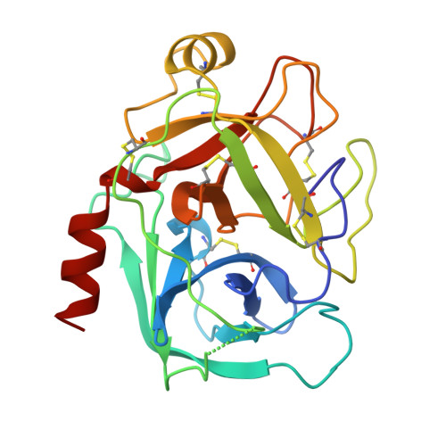
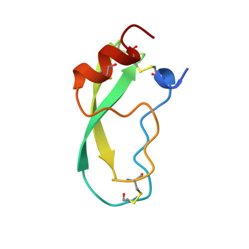
3BTW

THE CRYSTAL STRUCTURES OF THE COMPLEXES BETWEEN BOVINE BETA-TRYPSIN AND TEN P1 VARIANTS OF BPTI


Experimental Data Snapshot

  • Method: X-RAY DIFFRACTION
  • Resolution: 2.05 Å
  • R-Value Free: 0.238 
  • R-Value Work: 0.191 

Starting Model: experimental
View more details

wwPDB Validation   3D Report Full Report


This is version 1.5 of the entry. See complete history


Literature

The crystal structures of the complexes between bovine beta-trypsin and ten P1 variants of BPTI.

Helland, R.Otlewski, J.Sundheim, O.Dadlez, M.Smalas, A.O.

(1999) J Mol Biol 287: 923-942

  • DOI: https://doi.org/10.1006/jmbi.1999.2654
  • Primary Citation of Related Structures:  
    3BTD, 3BTE, 3BTF, 3BTG, 3BTH, 3BTK, 3BTM, 3BTQ, 3BTT, 3BTW

  • PubMed Abstract: 

    The high-resolution X-ray structures have been determined for ten complexes formed between bovine beta-trypsin and P1 variants (Gly, Asp, Glu, Gln, Thr, Met, Lys, His, Phe, Trp) of bovine pancreatic trypsin inhibitor (BPTI). All the complexes were crystallised from the same conditions. The structures of the P1 variants Asp, Glu, Gln and Thr, are reported here for the first time in complex with any serine proteinase. The resolution of the structures ranged from 1.75 to 2.05 A and the R-factors were about 19-20 %. The association constants of the mutants ranged from 1.5x10(4) to 1.7x10(13) M-1. All the structures could be fitted into well-defined electron density, and all had very similar global conformations. All the P1 mutant side-chains could be accomodated at the primary binding site, but relative to the P1 Lys, there were small local changes within the P1-S1 interaction site. These comprised: (1) changes in the number and dynamics of water molecules inside the pocket; (2) multiple conformations and non-optimal dihedral angles for some of the P1 side-chains, Ser190 and Gln192; and (3) changes in temperature factors of the pocket walls as well as the introduced P1 side-chain. Binding of the cognate P1 Lys is characterised by almost optimal dihedral angles, hydrogen bonding distances and angles, in addition to considerably lower temperature factors. Thus, the trypsin S1 pocket seems to be designed particularly for lysine binding.


  • Organizational Affiliation

    Department of Chemistry, University of Tromsø, Tromsø, 9037, Norway.


Macromolecules
Find similar proteins by:  (by identity cutoff)  |  3D Structure
Entity ID: 1
MoleculeChains Sequence LengthOrganismDetailsImage
PROTEIN (TRYPSIN)A [auth E]223Bos taurusMutation(s): 0 
EC: 3.4.21.4
UniProt
Find proteins for P00760 (Bos taurus)
Explore P00760 
Go to UniProtKB:  P00760
Entity Groups  
Sequence Clusters30% Identity50% Identity70% Identity90% Identity95% Identity100% Identity
UniProt GroupP00760
Sequence Annotations
Expand
  • Reference Sequence
Find similar proteins by:  (by identity cutoff)  |  3D Structure
Entity ID: 2
MoleculeChains Sequence LengthOrganismDetailsImage
PROTEIN (BOVINE PANCREATIC TRYPSIN INHIBITOR)B [auth I]58Bos taurusMutation(s): 2 
UniProt
Find proteins for P00974 (Bos taurus)
Explore P00974 
Go to UniProtKB:  P00974
Entity Groups  
Sequence Clusters30% Identity50% Identity70% Identity90% Identity95% Identity100% Identity
UniProt GroupP00974
Sequence Annotations
Expand
  • Reference Sequence
Experimental Data & Validation

Experimental Data

  • Method: X-RAY DIFFRACTION
  • Resolution: 2.05 Å
  • R-Value Free: 0.238 
  • R-Value Work: 0.191 
  • Space Group: I 2 2 2
Unit Cell:
Length ( Å )Angle ( ˚ )
a = 75.67α = 90
b = 84.78β = 90
c = 122.99γ = 90
Software Package:
Software NamePurpose
DENZOdata reduction
CCP4data reduction
X-PLORmodel building
X-PLORrefinement
CCP4data scaling
X-PLORphasing

Structure Validation

View Full Validation Report


Entry History 

Deposition Data

Revision History  (Full details and data files)

  • Version 1.0: 2000-03-13
    Type: Initial release
  • Version 1.1: 2008-04-26
    Changes: Version format compliance
  • Version 1.2: 2011-07-13
    Changes: Version format compliance
  • Version 1.3: 2018-03-14
    Changes: Database references
  • Version 1.4: 2023-08-30
    Changes: Data collection, Database references, Derived calculations, Refinement description
  • Version 1.5: 2024-11-20
    Changes: Structure summary