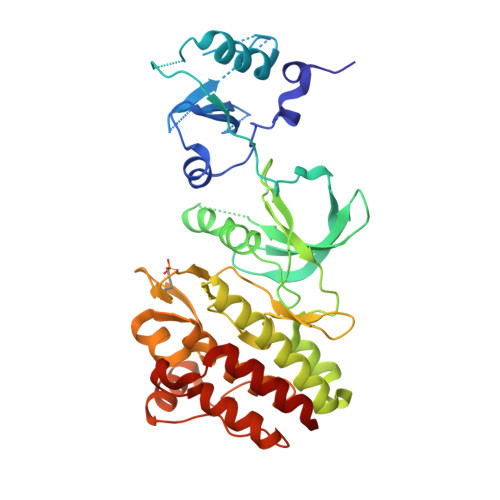
3CD3

Crystal structure of phosphorylated human feline sarcoma viral oncogene homologue (v-FES) in complex with staurosporine and a consensus peptide


Experimental Data Snapshot

  • Method: X-RAY DIFFRACTION
  • Resolution: 1.98 Å
  • R-Value Free: 0.247 
  • R-Value Work: 0.185 
  • R-Value Observed: 0.188 

Starting Model: experimental
View more details

wwPDB Validation   3D Report Full Report


Ligand Structure Quality Assessment 


This is version 1.4 of the entry. See complete history


Literature

Structural Coupling of SH2-Kinase Domains Links Fes and Abl Substrate Recognition and Kinase Activation

Filippakopoulos, P.Kofler, M.Hantschel, O.Gish, G.D.Grebien, F.Salah, E.Neudecker, P.Kay, L.E.Turk, B.E.Superti-Furga, G.Pawson, T.Knapp, S.

(2008) Cell 134: 793-803

  • DOI: https://doi.org/10.1016/j.cell.2008.07.047
  • Primary Citation of Related Structures:  
    3BKB, 3CBL, 3CD3

  • PubMed Abstract: 

    The SH2 domain of cytoplasmic tyrosine kinases can enhance catalytic activity and substrate recognition, but the molecular mechanisms by which this is achieved are poorly understood. We have solved the structure of the prototypic SH2-kinase unit of the human Fes tyrosine kinase, which appears specialized for positive signaling. In its active conformation, the SH2 domain tightly interacts with the kinase N-terminal lobe and positions the kinase alphaC helix in an active configuration through essential packing and electrostatic interactions. This interaction is stabilized by ligand binding to the SH2 domain. Our data indicate that Fes kinase activation is closely coupled to substrate recognition through cooperative SH2-kinase-substrate interactions. Similarly, we find that the SH2 domain of the active Abl kinase stimulates catalytic activity and substrate phosphorylation through a distinct SH2-kinase interface. Thus, the SH2 and catalytic domains of active Fes and Abl pro-oncogenic kinases form integrated structures essential for effective tyrosine kinase signaling.


  • Organizational Affiliation

    Structural Genomics Consortium, University of Oxford, Old Road Campus, Roosevelt Drive, Oxford OX3 7DQ, UK.


Macromolecules
Find similar proteins by:  (by identity cutoff)  |  3D Structure
Entity ID: 1
MoleculeChains Sequence LengthOrganismDetailsImage
Proto-oncogene tyrosine-protein kinase Fes/Fps377Homo sapiensMutation(s): 0 
Gene Names: FESFPS
EC: 2.7.10.2
UniProt & NIH Common Fund Data Resources
Find proteins for P07332 (Homo sapiens)
Explore P07332 
Go to UniProtKB:  P07332
PHAROS:  P07332
GTEx:  ENSG00000182511 
Entity Groups  
Sequence Clusters30% Identity50% Identity70% Identity90% Identity95% Identity100% Identity
UniProt GroupP07332
Sequence Annotations
Expand
  • Reference Sequence

Find similar proteins by:  Sequence   |   3D Structure  

Entity ID: 2
MoleculeChains Sequence LengthOrganismDetailsImage
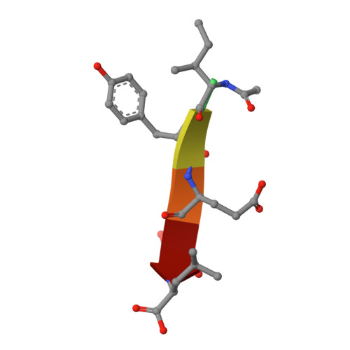
Synthetic peptide6N/AMutation(s): 0 
Sequence Annotations
Expand
  • Reference Sequence
Experimental Data & Validation

Experimental Data

  • Method: X-RAY DIFFRACTION
  • Resolution: 1.98 Å
  • R-Value Free: 0.247 
  • R-Value Work: 0.185 
  • R-Value Observed: 0.188 
  • Space Group: P 21 21 21
Unit Cell:
Length ( Å )Angle ( ˚ )
a = 35.451α = 90
b = 76.905β = 90
c = 150.632γ = 90
Software Package:
Software NamePurpose
SCALAdata scaling
PHASERphasing
REFMACrefinement
PDB_EXTRACTdata extraction
CrystalCleardata collection
MOSFLMdata reduction

Structure Validation

View Full Validation Report


Ligand Structure Quality Assessment 


Entry History 

Revision History  (Full details and data files)

  • Version 1.0: 2008-03-25
    Type: Initial release
  • Version 1.1: 2011-07-13
    Changes: Advisory, Version format compliance
  • Version 1.2: 2023-08-30
    Changes: Data collection, Database references, Derived calculations, Refinement description
  • Version 1.3: 2023-11-15
    Changes: Data collection
  • Version 1.4: 2024-11-06
    Changes: Structure summary