3DZ7

Human AdoMetDC with 5'-[(carboxamidomethyl)methylamino]-5'-deoxy-8-methyladenosine


Experimental Data Snapshot

  • Method: X-RAY DIFFRACTION
  • Resolution: 1.91 Å
  • R-Value Free: 0.208 
  • R-Value Work: 0.197 
  • R-Value Observed: 0.197 

Starting Model: experimental
View more details

wwPDB Validation   3D Report Full Report


Ligand Structure Quality Assessment 


This is version 2.0 of the entry. See complete history


Literature

New Insights into the Design of Inhibitors of Human S-Adenosylmethionine Decarboxylase: Studies of Adenine C8 Substitution in Structural Analogues of S-Adenosylmethionine

McCloskey, D.E.Bale, S.Secrist III, J.A.Tiwari, A.Moss III, T.H.Valiyaveettil, J.Brooks, W.H.Guida, W.C.Pegg, A.E.Ealick, S.E.

(2009) J Med Chem 52: 1388-1407

  • DOI: https://doi.org/10.1021/jm801126a
  • Primary Citation of Related Structures:  
    3DZ2, 3DZ3, 3DZ4, 3DZ5, 3DZ6, 3DZ7

  • PubMed Abstract: 

    S-adenosylmethionine decarboxylase (AdoMetDC) is a critical enzyme in the polyamine biosynthetic pathway and depends on a pyruvoyl group for the decarboxylation process. The crystal structures of the enzyme with various inhibitors at the active site have shown that the adenine base of the ligands adopts an unusual syn conformation when bound to the enzyme. To determine whether compounds that favor the syn conformation in solution would be more potent AdoMetDC inhibitors, several series of AdoMet substrate analogues with a variety of substituents at the 8-position of adenine were synthesized and analyzed for their ability to inhibit hAdoMetDC. The biochemical analysis indicated that an 8-methyl substituent resulted in more potent inhibitors, yet most other 8-substitutions provided no benefit over the parent compound. To understand these results, we used computational modeling and X-ray crystallography to study C(8)-substituted adenine analogues bound in the active site.


  • Organizational Affiliation

    Department of Cellular and Molecular Physiology, Pennsylvania State University College of Medicine, Hershey, Pennsylvania 17033, USA.


Macromolecules
Find similar proteins by:  (by identity cutoff)  |  3D Structure
Entity ID: 1
MoleculeChains Sequence LengthOrganismDetailsImage
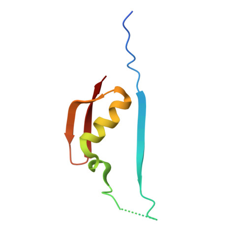
S-adenosylmethionine decarboxylase beta chainA [auth B]67Homo sapiensMutation(s): 0 
Gene Names: AMD1AMD
EC: 4.1.1.50
UniProt & NIH Common Fund Data Resources
Find proteins for P17707 (Homo sapiens)
Explore P17707 
Go to UniProtKB:  P17707
PHAROS:  P17707
GTEx:  ENSG00000123505 
Entity Groups  
Sequence Clusters30% Identity50% Identity70% Identity90% Identity95% Identity100% Identity
UniProt GroupP17707
Sequence Annotations
Expand
  • Reference Sequence
Find similar proteins by:  (by identity cutoff)  |  3D Structure
Entity ID: 2
MoleculeChains Sequence LengthOrganismDetailsImage
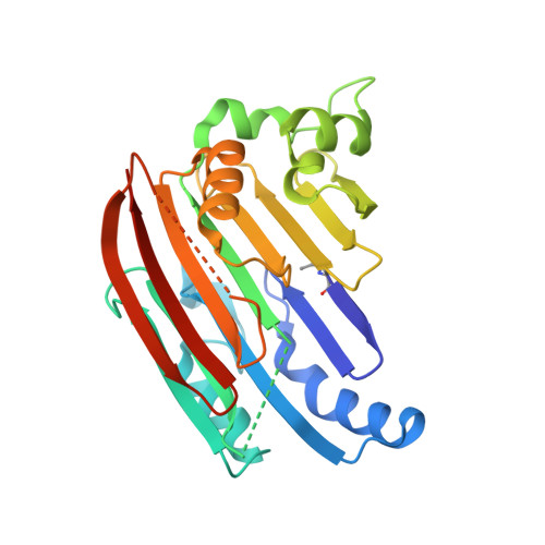
S-adenosylmethionine decarboxylase alpha chainB [auth A]267Homo sapiensMutation(s): 0 
Gene Names: AMD1AMD
EC: 4.1.1.50
UniProt & NIH Common Fund Data Resources
Find proteins for P17707 (Homo sapiens)
Explore P17707 
Go to UniProtKB:  P17707
PHAROS:  P17707
GTEx:  ENSG00000123505 
Entity Groups  
Sequence Clusters30% Identity50% Identity70% Identity90% Identity95% Identity100% Identity
UniProt GroupP17707
Sequence Annotations
Expand
  • Reference Sequence
Small Molecules
Ligands 2 Unique
IDChains Name / Formula / InChI Key2D Diagram3D Interactions
O8M
Query on O8M

Download Ideal Coordinates CCD File 
D [auth A]2-[{[(2R,3S,4R,5R)-5-(6-amino-8-methyl-9H-purin-9-yl)-3,4-dihydroxytetrahydrofuran-2-yl]methyl}(methyl)amino]acetamide
C14 H21 N7 O4
IRWBMXVMEDHEJW-FRJWGUMJSA-N
PUT
Query on PUT

Download Ideal Coordinates CCD File 
C [auth B]1,4-DIAMINOBUTANE
C4 H12 N2
KIDHWZJUCRJVML-UHFFFAOYSA-N
Experimental Data & Validation

Experimental Data

  • Method: X-RAY DIFFRACTION
  • Resolution: 1.91 Å
  • R-Value Free: 0.208 
  • R-Value Work: 0.197 
  • R-Value Observed: 0.197 
  • Space Group: C 1 2 1
Unit Cell:
Length ( Å )Angle ( ˚ )
a = 99.646α = 90
b = 50.752β = 105.34
c = 68.9γ = 90
Software Package:
Software NamePurpose
DENZOdata reduction
SCALEPACKdata scaling
CNSrefinement
PDB_EXTRACTdata extraction
ADSCdata collection
HKL-2000data reduction
CNSphasing

Structure Validation

View Full Validation Report


Ligand Structure Quality Assessment 


Entry History 

Deposition Data

Revision History  (Full details and data files)

  • Version 1.0: 2009-03-10
    Type: Initial release
  • Version 1.1: 2011-07-13
    Changes: Version format compliance
  • Version 1.2: 2017-10-25
    Changes: Advisory, Refinement description
  • Version 1.3: 2023-08-30
    Changes: Advisory, Data collection, Database references, Derived calculations, Refinement description
  • Version 2.0: 2023-11-15
    Changes: Atomic model, Data collection, Derived calculations