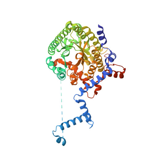
3E2R

Crystal Structure PutA86-630 Mutant Y540S Complexed with L-tetrahydro-2-furoic acid


Experimental Data Snapshot

  • Method: X-RAY DIFFRACTION
  • Resolution: 1.85 Å
  • R-Value Free: 0.227 
  • R-Value Work: 0.198 
  • R-Value Observed: 0.199 

wwPDB Validation   3D Report Full Report


Ligand Structure Quality Assessment 


This is version 1.4 of the entry. See complete history


Literature

A conserved active site tyrosine residue of proline dehydrogenase helps enforce the preference for proline over hydroxyproline as the substrate.

Ostrander, E.L.Larson, J.D.Schuermann, J.P.Tanner, J.J.

(2009) Biochemistry 48: 951-959

  • DOI: https://doi.org/10.1021/bi802094k
  • Primary Citation of Related Structures:  
    3E2Q, 3E2R, 3E2S

  • PubMed Abstract: 

    Proline dehydrogenase (PRODH) catalyzes the oxidation of l-proline to Delta-1-pyrroline-5-carboxylate. PRODHs exhibit a pronounced preference for proline over hydroxyproline (trans-4-hydroxy-l-proline) as the substrate, but the basis for specificity is unknown. The goal of this study, therefore, is to gain insight into the structural determinants of substrate specificity of this class of enzyme, with a focus on understanding how PRODHs discriminate between the two closely related molecules, proline and hydroxyproline. Two site-directed mutants of the PRODH domain of Escherichia coli PutA were created: Y540A and Y540S. Kinetics measurements were performed with both mutants. Crystal structures of Y540S complexed with hydroxyproline, proline, and the proline analogue l-tetrahydro-2-furoic acid were determined at resolutions of 1.75, 1.90, and 1.85 A, respectively. Mutation of Tyr540 increases the catalytic efficiency for hydroxyproline 3-fold and decreases the specificity for proline by factors of 20 (Y540S) and 50 (Y540A). The structures show that removal of the large phenol side chain increases the volume of the substrate-binding pocket, allowing sufficient room for the 4-hydroxyl of hydroxyproline. Furthermore, the introduced serine residue participates in recognition of hydroxyproline by forming a hydrogen bond with the 4-hydroxyl. This result has implications for understanding the substrate specificity of the related enzyme human hydroxyproline dehydrogenase, which has serine in place of tyrosine at this key active site position. The kinetic and structural results suggest that Tyr540 is an important determinant of specificity. Structurally, it serves as a negative filter for hydroxyproline by clashing with the 4-hydroxyl group of this potential substrate.


  • Organizational Affiliation

    Department of Chemistry, University of Missouri, Columbia, Missouri 65211, USA.


Macromolecules
Find similar proteins by:  (by identity cutoff)  |  3D Structure
Entity ID: 1
MoleculeChains Sequence LengthOrganismDetailsImage
Proline dehydrogenase551Escherichia coli K-12Mutation(s): 1 
Gene Names: putApoaA
EC: 1.5.99.8 (PDB Primary Data), 1.2.1.88 (UniProt), 1.5.5.2 (UniProt)
UniProt
Find proteins for P09546 (Escherichia coli (strain K12))
Explore P09546 
Go to UniProtKB:  P09546
Entity Groups  
Sequence Clusters30% Identity50% Identity70% Identity90% Identity95% Identity100% Identity
UniProt GroupP09546
Sequence Annotations
Expand
  • Reference Sequence
Small Molecules
Ligands 4 Unique
IDChains Name / Formula / InChI Key2D Diagram3D Interactions
FAD
Query on FAD

Download Ideal Coordinates CCD File 
B [auth A]FLAVIN-ADENINE DINUCLEOTIDE
C27 H33 N9 O15 P2
VWWQXMAJTJZDQX-UYBVJOGSSA-N
1PE
Query on 1PE

Download Ideal Coordinates CCD File 
E [auth A],
F [auth A]
PENTAETHYLENE GLYCOL
C10 H22 O6
JLFNLZLINWHATN-UHFFFAOYSA-N
CIT
Query on CIT

Download Ideal Coordinates CCD File 
D [auth A]CITRIC ACID
C6 H8 O7
KRKNYBCHXYNGOX-UHFFFAOYSA-N
TFB
Query on TFB

Download Ideal Coordinates CCD File 
C [auth A]TETRAHYDROFURAN-2-CARBOXYLIC ACID
C5 H8 O3
UJJLJRQIPMGXEZ-BYPYZUCNSA-N
Experimental Data & Validation

Experimental Data

  • Method: X-RAY DIFFRACTION
  • Resolution: 1.85 Å
  • R-Value Free: 0.227 
  • R-Value Work: 0.198 
  • R-Value Observed: 0.199 
  • Space Group: I 2 2 2
Unit Cell:
Length ( Å )Angle ( ˚ )
a = 72.648α = 90
b = 140.102β = 90
c = 146.665γ = 90
Software Package:
Software NamePurpose
DENZOdata reduction
SCALEPACKdata scaling
PHENIXrefinement
PDB_EXTRACTdata extraction
HKL-2000data reduction
HKL-2000data scaling

Structure Validation

View Full Validation Report


Ligand Structure Quality Assessment 


Entry History 

Deposition Data

  • Released Date: 2009-02-03 
  • Deposition Author(s): Tanner, J.J.

Revision History  (Full details and data files)

  • Version 1.0: 2009-02-03
    Type: Initial release
  • Version 1.1: 2011-07-13
    Changes: Version format compliance
  • Version 1.2: 2017-10-25
    Changes: Refinement description
  • Version 1.3: 2021-10-20
    Changes: Database references, Derived calculations
  • Version 1.4: 2024-02-21
    Changes: Data collection