3EBP

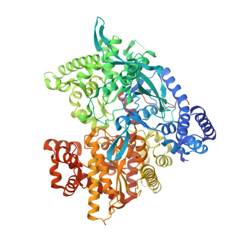
Glycogen Phosphorylase b/flavopiridol complex


Experimental Data Snapshot

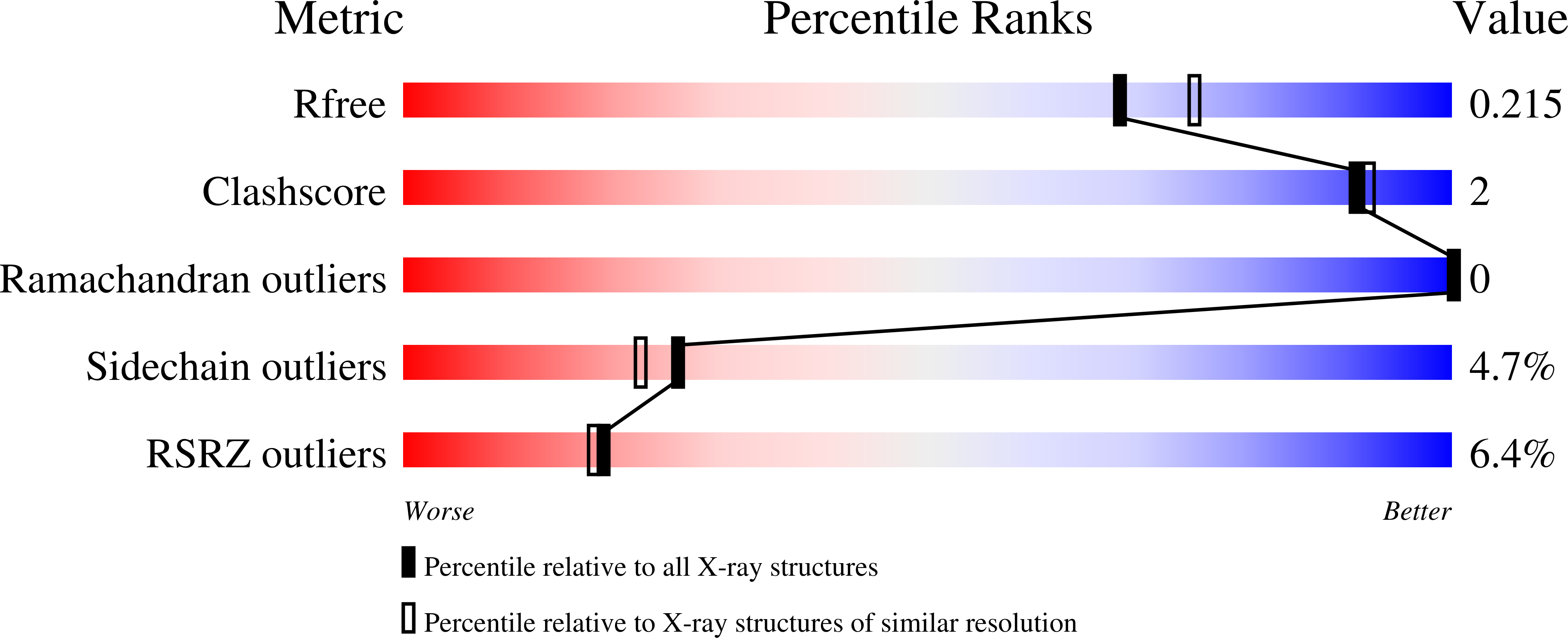
  • Method: X-RAY DIFFRACTION
  • Resolution: 2.00 Å
  • R-Value Free: 0.215 
  • R-Value Work: 0.188 
  • R-Value Observed: 0.190 

Starting Model: experimental
View more details

wwPDB Validation   3D Report Full Report


Ligand Structure Quality Assessment 


This is version 1.5 of the entry. See complete history


Literature

Sourcing the affinity of flavonoids for the glycogen phosphorylase inhibitor site via crystallography, kinetics and QM/MM-PBSA binding studies: Comparison of chrysin and flavopiridol

Tsitsanou, K.E.Hayes, J.M.Keramioti, M.Mamais, M.Oikonomakos, N.G.Kato, A.Leonidas, D.D.Zographos, S.E.

(2013) Food Chem Toxicol 61: 14-27

  • DOI: https://doi.org/10.1016/j.fct.2012.12.030
  • Primary Citation of Related Structures:  
    3EBO, 3EBP

  • PubMed Abstract: 

    Flavonoids have been discovered as novel inhibitors of glycogen phosphorylase (GP), a target to control hyperglycemia in type 2 diabetes. To elucidate the mechanism of inhibition, we have determined the crystal structure of the GPb-chrysin complex at 1.9 Å resolution. Chrysin is accommodated at the inhibitor site intercalating between the aromatic side chains of Phe285 and Tyr613 through π-stacking interactions. Chrysin binds to GPb approximately 15 times weaker (Ki=19.01 μM) than flavopiridol (Ki=1.24 μM), exclusively at the inhibitor site, and both inhibitors display similar behavior with respect to AMP. To identify the source of flavopiridols' stronger affinity, molecular docking with Glide and postdocking binding free energy calculations using QM/MM-PBSA have been performed and compared. Whereas docking failed to correctly rank inhibitor binding conformations, the QM/MM-PBSA method employing M06-2X/6-31+G to model the π-stacking interactions correctly reproduced the experimental results. Flavopiridols' greater binding affinity is sourced to favorable interactions of the cationic 4-hydroxypiperidin-1-yl substituent with GPb, with desolvation effects limited by the substituent conformation adopted in the crystallographic complex. Further successful predictions using QM/MM-PBSA for the flavonoid quercetagetin (which binds at the allosteric site) leads us to propose the methodology as a useful and inexpensive tool to predict flavonoid binding.


  • Organizational Affiliation

    Institute of Biology, Medicinal Chemistry and Biotechnology, National Hellenic Research Foundation, 48 Vassileos Constantinou Avenue, Athens 11635, Greece.


Macromolecules
Find similar proteins by:  (by identity cutoff)  |  3D Structure
Entity ID: 1
MoleculeChains Sequence LengthOrganismDetailsImage
Glycogen phosphorylase, muscle form842Oryctolagus cuniculusMutation(s): 0 
EC: 2.4.1.1
UniProt
Find proteins for P00489 (Oryctolagus cuniculus)
Explore P00489 
Go to UniProtKB:  P00489
Entity Groups  
Sequence Clusters30% Identity50% Identity70% Identity90% Identity95% Identity100% Identity
UniProt GroupP00489
Sequence Annotations
Expand
  • Reference Sequence
Small Molecules
Ligands 1 Unique
IDChains Name / Formula / InChI Key2D Diagram3D Interactions
CPB
Query on CPB

Download Ideal Coordinates CCD File 
B [auth A]2-(2-CHLORO-PHENYL)-5,7-DIHYDROXY-8-(3-HYDROXY-1-METHYL-PIPERIDIN-4-YL)-4H-BENZOPYRAN-4-ONE
C21 H20 Cl N O5
BIIVYFLTOXDAOV-YVEFUNNKSA-N
Modified Residues  1 Unique
IDChains TypeFormula2D DiagramParent
LLP
Query on LLP
A
L-PEPTIDE LINKINGC14 H22 N3 O7 PLYS
Binding Affinity Annotations 
IDSourceBinding Affinity
CPB PDBBind:  3EBP Ki: 1240 (nM) from 1 assay(s)
BindingDB:  3EBP IC50: min: 1000, max: 1200 (nM) from 2 assay(s)
Experimental Data & Validation

Experimental Data

  • Method: X-RAY DIFFRACTION
  • Resolution: 2.00 Å
  • R-Value Free: 0.215 
  • R-Value Work: 0.188 
  • R-Value Observed: 0.190 
  • Space Group: P 43 21 2
Unit Cell:
Length ( Å )Angle ( ˚ )
a = 128.653α = 90
b = 128.653β = 90
c = 116.489γ = 90
Software Package:
Software NamePurpose
MAR345dtbdata collection
REFMACrefinement
DENZOdata reduction
SCALEPACKdata scaling
REFMACphasing

Structure Validation

View Full Validation Report


Ligand Structure Quality Assessment 


Entry History 

Deposition Data

Revision History  (Full details and data files)

  • Version 1.0: 2009-09-01
    Type: Initial release
  • Version 1.1: 2011-07-13
    Changes: Version format compliance
  • Version 1.2: 2013-01-16
    Changes: Database references, Structure summary
  • Version 1.3: 2013-12-11
    Changes: Database references
  • Version 1.4: 2023-11-01
    Changes: Data collection, Database references, Derived calculations, Refinement description
  • Version 1.5: 2023-11-15
    Changes: Data collection