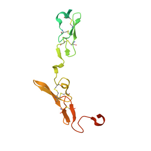
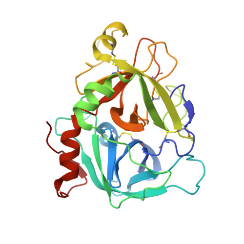
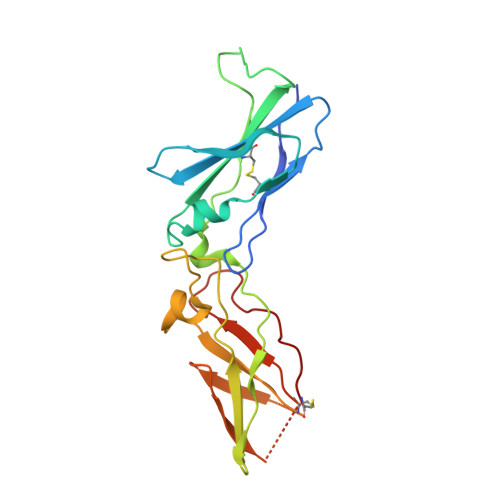
3ELA

Crystal structure of active site inhibited coagulation factor VIIA mutant in complex with soluble tissue factor


Experimental Data Snapshot

  • Method: X-RAY DIFFRACTION
  • Resolution: 2.20 Å
  • R-Value Free: 0.294 
  • R-Value Work: 0.233 
  • R-Value Observed: 0.236 

Starting Model: experimental
View more details

wwPDB Validation   3D Report Full Report


Ligand Structure Quality Assessment 


This is version 1.7 of the entry. See complete history


Literature

Mechanism of the Ca2+-induced enhancement of the intrinsic factor VIIa activity

Bjelke, J.R.Olsen, O.H.Fodje, M.Svensson, L.A.Bang, S.Bolt, G.Kragelund, B.B.Persson, E.

(2008) J Biol Chem 283: 25863-25870

  • DOI: https://doi.org/10.1074/jbc.M800841200
  • Primary Citation of Related Structures:  
    3ELA

  • PubMed Abstract: 

    The intrinsic activity of coagulation factor VIIa (FVIIa) is dependent on Ca(2+) binding to a loop (residues 210-220) in the protease domain. Structural analysis revealed that Ca(2+) may enhance the activity by attenuating electrostatic repulsion of Glu(296) and/or by facilitating interactions between the loop and Lys(161) in the N-terminal tail. In support of the first mechanism, the mutations E296V and D212N resulted in similar, about 2-fold, enhancements of the amidolytic activity. Moreover, mutation of the Lys(161)-interactive residue Asp(217) or Asp(219) to Ala reduced the amidolytic activity by 40-50%, whereas the K161A mutation resulted in 80% reduction. Hence one of these Asp residues in the Ca(2+)-binding loop appears to suffice for some residual interaction with Lys(161), whereas the more severe effect upon replacement of Lys(161) is due to abrogation of the interaction with the N-terminal tail. However, Ca(2+) attenuation of the repulsion between Asp(212) and Glu(296) keeps the activity above that of apoFVIIa. Altogether, our data suggest that repulsion involving Asp(212) in the Ca(2+)-binding loop suppresses FVIIa activity and that optimal activity requires a favorable interaction between the Ca(2+)-binding loop and the N-terminal tail. Crystal structures of tissue factor-bound FVIIa(D212N) and FVIIa(V158D/E296V/M298Q) revealed altered hydrogen bond networks, resembling those in factor Xa and thrombin, after introduction of the D212N and E296V mutations plausibly responsible for tethering the N-terminal tail to the activation domain. The charge repulsion between the Ca(2+)-binding loop and the activation domain appeared to be either relieved by charge removal and new hydrogen bonds (D212N) or abolished (E296V). We propose that Ca(2+) stimulates the intrinsic FVIIa activity by a combination of charge neutralization and loop stabilization.


  • Organizational Affiliation

    Department of Protein Structure and Biophysics, Novo Nordisk A/S, Novo Nordisk Park, DK-2760 Måløv, Denmark.


Macromolecules
Find similar proteins by:  (by identity cutoff)  |  3D Structure
Entity ID: 1
MoleculeChains Sequence LengthOrganismDetailsImage
Coagulation factor VIIA light chainA [auth L]152Homo sapiensMutation(s): 0 
Gene Names: F7
EC: 3.4.21.21
UniProt & NIH Common Fund Data Resources
Find proteins for P08709 (Homo sapiens)
Explore P08709 
Go to UniProtKB:  P08709
PHAROS:  P08709
GTEx:  ENSG00000057593 
Entity Groups  
Sequence Clusters30% Identity50% Identity70% Identity90% Identity95% Identity100% Identity
UniProt GroupP08709
Sequence Annotations
Expand
  • Reference Sequence
Find similar proteins by:  (by identity cutoff)  |  3D Structure
Entity ID: 2
MoleculeChains Sequence LengthOrganismDetailsImage
Coagulation factor VIIA heavy chainB [auth H]254Homo sapiensMutation(s): 3 
Gene Names: F7
EC: 3.4.21.21
UniProt & NIH Common Fund Data Resources
Find proteins for P08709 (Homo sapiens)
Explore P08709 
Go to UniProtKB:  P08709
PHAROS:  P08709
GTEx:  ENSG00000057593 
Entity Groups  
Sequence Clusters30% Identity50% Identity70% Identity90% Identity95% Identity100% Identity
UniProt GroupP08709
Sequence Annotations
Expand
  • Reference Sequence
Find similar proteins by:  (by identity cutoff)  |  3D Structure
Entity ID: 3
MoleculeChains Sequence LengthOrganismDetailsImage
Tissue factorC [auth T]209Homo sapiensMutation(s): 0 
Gene Names: F3
UniProt & NIH Common Fund Data Resources
Find proteins for P13726 (Homo sapiens)
Explore P13726 
Go to UniProtKB:  P13726
PHAROS:  P13726
GTEx:  ENSG00000117525 
Entity Groups  
Sequence Clusters30% Identity50% Identity70% Identity90% Identity95% Identity100% Identity
UniProt GroupP13726
Sequence Annotations
Expand
  • Reference Sequence
Biologically Interesting Molecules (External Reference) 1 Unique
Experimental Data & Validation

Experimental Data

  • Method: X-RAY DIFFRACTION
  • Resolution: 2.20 Å
  • R-Value Free: 0.294 
  • R-Value Work: 0.233 
  • R-Value Observed: 0.236 
  • Space Group: P 1 21 1
Unit Cell:
Length ( Å )Angle ( ˚ )
a = 78.361α = 90
b = 68.558β = 90.22
c = 78.817γ = 90
Software Package:
Software NamePurpose
MAR345data collection
CCP4model building
MOLREPphasing
REFMACrefinement
XDSdata reduction
XDSdata scaling
CCP4phasing

Structure Validation

View Full Validation Report


Ligand Structure Quality Assessment 


Entry History 

Deposition Data

Revision History  (Full details and data files)

  • Version 1.0: 2008-11-04
    Type: Initial release
  • Version 1.1: 2011-07-13
    Changes: Atomic model, Database references, Derived calculations, Non-polymer description, Structure summary, Version format compliance
  • Version 1.2: 2013-02-27
    Changes: Other
  • Version 1.3: 2017-10-25
    Changes: Data collection, Refinement description
  • Version 1.4: 2020-07-29
    Type: Remediation
    Reason: Carbohydrate remediation
    Changes: Data collection, Derived calculations, Structure summary
  • Version 1.5: 2021-11-10
    Changes: Database references, Structure summary
  • Version 1.6: 2023-11-01
    Changes: Data collection, Refinement description
  • Version 1.7: 2024-10-30
    Changes: Structure summary