3G5K

Structure and activity of human mitochondrial peptide deformylase, a novel cancer target


Experimental Data Snapshot

  • Method: X-RAY DIFFRACTION
  • Resolution: 1.70 Å
  • R-Value Free: 0.193 
  • R-Value Work: 0.164 
  • R-Value Observed: 0.165 

Starting Model: experimental
View more details

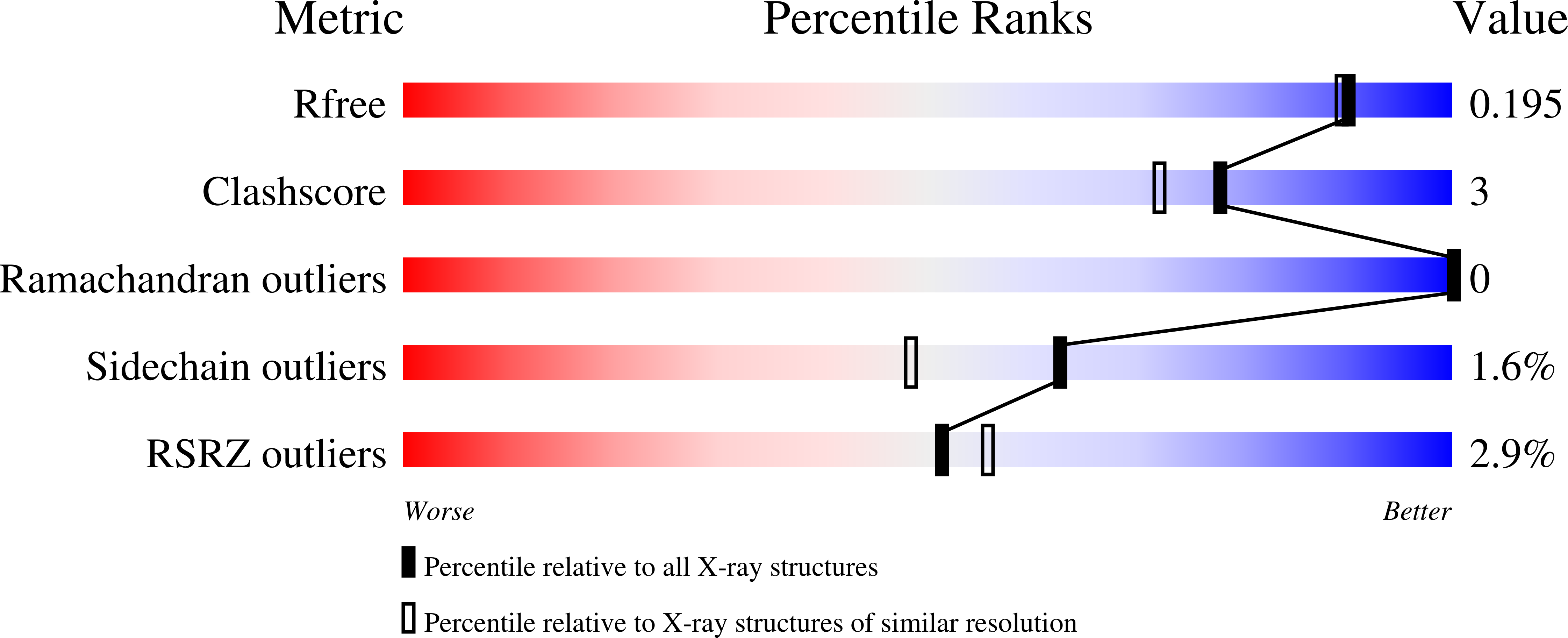
wwPDB Validation   3D Report Full Report


Ligand Structure Quality Assessment 


This is version 1.2 of the entry. See complete history


Literature

Structure and activity of human mitochondrial peptide deformylase, a novel cancer target

Escobar-Alvarez, S.Goldgur, Y.Yang, G.Ouerfelli, O.Li, Y.Scheinberg, D.A.

(2009) J Mol Biol 387: 1211-1228

  • DOI: https://doi.org/10.1016/j.jmb.2009.02.032
  • Primary Citation of Related Structures:  
    3G5K, 3G5P

  • PubMed Abstract: 

    Peptide deformylase proteins (PDFs) participate in the N-terminal methionine excision pathway of newly synthesized peptides. We show that the human PDF (HsPDF) can deformylate its putative substrates derived from mitochondrial DNA-encoded proteins. The first structural model of a mammalian PDF (1.7 A), HsPDF, shows a dimer with conserved topology of the catalytic residues and fold as non-mammalian PDFs. The HsPDF C-terminus topology and the presence of a helical loop (H2 and H3), however, shape a characteristic active site entrance. The structure of HsPDF bound to the peptidomimetic inhibitor actinonin (1.7 A) identified the substrate-binding site. A defined S1' pocket, but no S2' or S3' substrate-binding pockets, exists. A conservation of PDF-actinonin interaction across PDFs was observed. Despite the lack of true S2' and S3' binding pockets, confirmed through peptide binding modeling, enzyme kinetics suggest a combined contribution from P2'and P3' positions of a formylated peptide substrate to turnover.


  • Organizational Affiliation

    Molecular Pharmacology and Chemistry Program, Sloan-Kettering Institute, 415 E. 68th Street Zuckerman Z1941, New York, NY 10065, USA.


Macromolecules
Find similar proteins by:  (by identity cutoff)  |  3D Structure
Entity ID: 1
MoleculeChains Sequence LengthOrganismDetailsImage
Peptide deformylase, mitochondrial
A, B, C, D
183Homo sapiensMutation(s): 0 
Gene Names: PDFPDF1A
EC: 3.5.1.88
UniProt & NIH Common Fund Data Resources
Find proteins for Q9HBH1 (Homo sapiens)
Explore Q9HBH1 
Go to UniProtKB:  Q9HBH1
GTEx:  ENSG00000258429 
Entity Groups  
Sequence Clusters30% Identity50% Identity70% Identity90% Identity95% Identity100% Identity
UniProt GroupQ9HBH1
Sequence Annotations
Expand
  • Reference Sequence
Small Molecules
Ligands 2 Unique
IDChains Name / Formula / InChI Key2D Diagram3D Interactions
BB2
Query on BB2

Download Ideal Coordinates CCD File 
E [auth A],
G [auth B],
I [auth C],
K [auth D]
ACTINONIN
C19 H35 N3 O5
XJLATMLVMSFZBN-VYDXJSESSA-N
CO
Query on CO

Download Ideal Coordinates CCD File 
F [auth A],
H [auth B],
J [auth C],
L [auth D]
COBALT (II) ION
Co
XLJKHNWPARRRJB-UHFFFAOYSA-N
Binding Affinity Annotations 
IDSourceBinding Affinity
BB2 PDBBind:  3G5K Ki: 0.3 (nM) from 1 assay(s)
Experimental Data & Validation

Experimental Data

  • Method: X-RAY DIFFRACTION
  • Resolution: 1.70 Å
  • R-Value Free: 0.193 
  • R-Value Work: 0.164 
  • R-Value Observed: 0.165 
  • Space Group: C 1 2 1
Unit Cell:
Length ( Å )Angle ( ˚ )
a = 116.144α = 90
b = 77.83β = 107.46
c = 110.532γ = 90
Software Package:
Software NamePurpose
ADSCdata collection
AMoREphasing
REFMACrefinement
HKL-2000data reduction
HKL-2000data scaling

Structure Validation

View Full Validation Report


Ligand Structure Quality Assessment 


Entry History 

Deposition Data

Revision History  (Full details and data files)

  • Version 1.0: 2009-04-07
    Type: Initial release
  • Version 1.1: 2011-07-13
    Changes: Advisory, Refinement description, Version format compliance
  • Version 1.2: 2023-09-06
    Changes: Data collection, Database references, Derived calculations, Refinement description