3GW9

Crystal structure of sterol 14-alpha demethylase (CYP51) from Trypanosoma brucei bound to an inhibitor N-(1-(2,4-dichlorophenyl)-2-(1H-imidazol-1-yl)ethyl)-4-(5-phenyl-1,3,4-oxaziazol-2-yl)benzamide


Experimental Data Snapshot

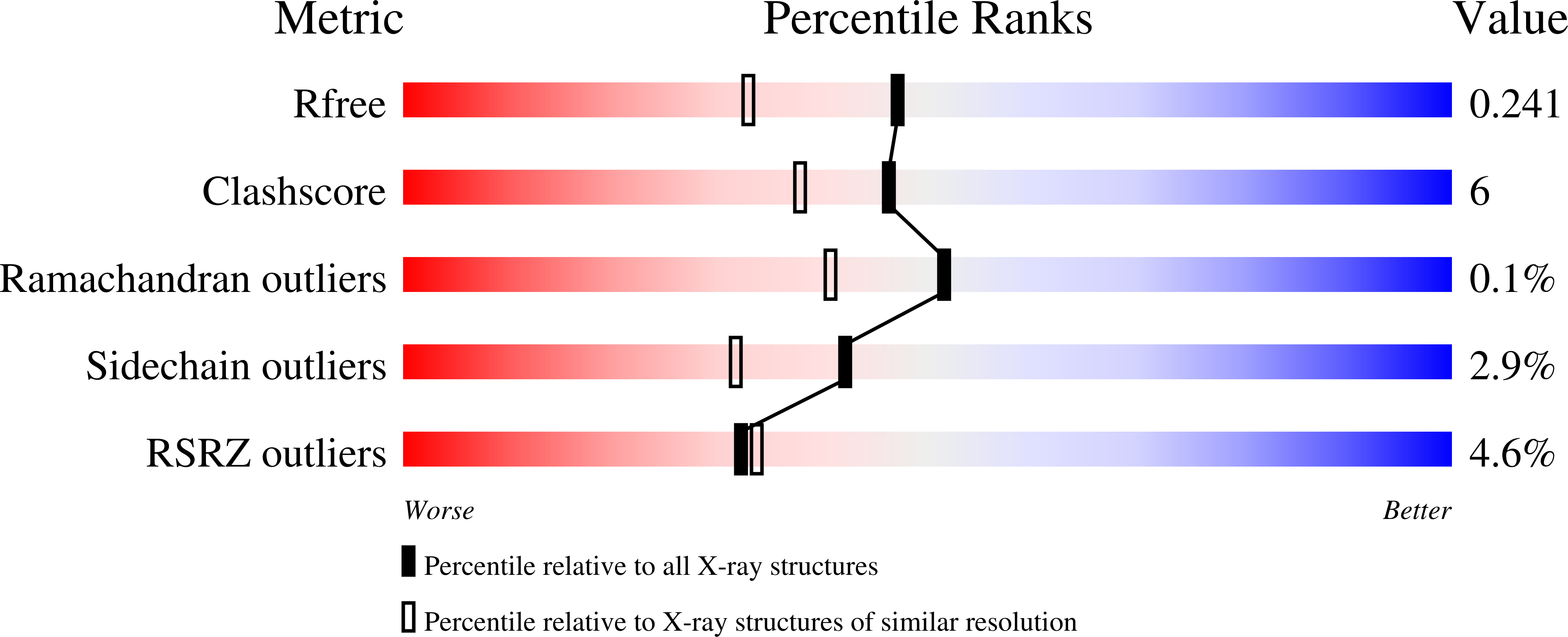
  • Method: X-RAY DIFFRACTION
  • Resolution: 1.87 Å
  • R-Value Free: 0.238 
  • R-Value Work: 0.189 
  • R-Value Observed: 0.191 

Starting Model: experimental
View more details

wwPDB Validation   3D Report Full Report


Ligand Structure Quality Assessment 


This is version 1.3 of the entry. See complete history


Literature

Crystal structures of Trypanosoma brucei sterol 14alpha-demethylase and implications for selective treatment of human infections.

Lepesheva, G.I.Park, H.W.Hargrove, T.Y.Vanhollebeke, B.Wawrzak, Z.Harp, J.M.Sundaramoorthy, M.Nes, W.D.Pays, E.Chaudhuri, M.Villalta, F.Waterman, M.R.

(2010) J Biol Chem 285: 1773-1780

  • DOI: https://doi.org/10.1074/jbc.M109.067470
  • Primary Citation of Related Structures:  
    3G1Q, 3GW9

  • PubMed Abstract: 

    Sterol 14alpha-demethylase (14DM, the CYP51 family of cytochrome P450) is an essential enzyme in sterol biosynthesis in eukaryotes. It serves as a major drug target for fungal diseases and can potentially become a target for treatment of human infections with protozoa. Here we present 1.9 A resolution crystal structures of 14DM from the protozoan pathogen Trypanosoma brucei, ligand-free and complexed with a strong chemically selected inhibitor N-1-(2,4-dichlorophenyl)-2-(1H-imidazol-1-yl)ethyl)-4-(5-phenyl-1,3,4-oxadi-azol-2-yl)benzamide that we previously found to produce potent antiparasitic effects in Trypanosomatidae. This is the first structure of a eukaryotic microsomal 14DM that acts on sterol biosynthesis, and it differs profoundly from that of the water-soluble CYP51 family member from Mycobacterium tuberculosis, both in organization of the active site cavity and in the substrate access channel location. Inhibitor binding does not cause large scale conformational rearrangements, yet induces unanticipated local alterations in the active site, including formation of a hydrogen bond network that connects, via the inhibitor amide group fragment, two remote functionally essential protein segments and alters the heme environment. The inhibitor binding mode provides a possible explanation for both its functionally irreversible effect on the enzyme activity and its selectivity toward the 14DM from human pathogens versus the human 14DM ortholog. The structures shed new light on 14DM functional conservation and open an excellent opportunity for directed design of novel antiparasitic drugs.


  • Organizational Affiliation

    Department of Biochemistry, Vanderbilt University, Nashville, Tennessee 37232, USA. [email protected]


Macromolecules
Find similar proteins by:  (by identity cutoff)  |  3D Structure
Entity ID: 1
MoleculeChains Sequence LengthOrganismDetailsImage
STEROL 14ALPHA-DEMETHYLASE
A, B, C, D
450Trypanosoma bruceiMutation(s): 4 
Gene Names: CYP51Tb11.02.4080
EC: 1.14.13.70
UniProt
Find proteins for Q385E8 (Trypanosoma brucei brucei (strain 927/4 GUTat10.1))
Explore Q385E8 
Go to UniProtKB:  Q385E8
Entity Groups  
Sequence Clusters30% Identity50% Identity70% Identity90% Identity95% Identity100% Identity
UniProt GroupQ385E8
Sequence Annotations
Expand
  • Reference Sequence
Small Molecules
Ligands 2 Unique
IDChains Name / Formula / InChI Key2D Diagram3D Interactions
HEM
Query on HEM

Download Ideal Coordinates CCD File 
E [auth A],
G [auth B],
I [auth C],
K [auth D]
PROTOPORPHYRIN IX CONTAINING FE
C34 H32 Fe N4 O4
KABFMIBPWCXCRK-RGGAHWMASA-L
VNI
Query on VNI

Download Ideal Coordinates CCD File 
F [auth A],
H [auth B],
J [auth C],
L [auth D]
N-[(1R)-1-(2,4-dichlorophenyl)-2-(1H-imidazol-1-yl)ethyl]-4-(5-phenyl-1,3,4-oxadiazol-2-yl)benzamide
C26 H19 Cl2 N5 O2
CJPLMXOWZZCYHJ-QHCPKHFHSA-N
Binding Affinity Annotations 
IDSourceBinding Affinity
VNI BindingDB:  3GW9 Kd: min: 8.3, max: 15 (nM) from 2 assay(s)
Experimental Data & Validation

Experimental Data

  • Method: X-RAY DIFFRACTION
  • Resolution: 1.87 Å
  • R-Value Free: 0.238 
  • R-Value Work: 0.189 
  • R-Value Observed: 0.191 
  • Space Group: P 1
Unit Cell:
Length ( Å )Angle ( ˚ )
a = 60.078α = 74.74
b = 79.113β = 79.13
c = 115.998γ = 68.57
Software Package:
Software NamePurpose
HKL-2000data collection
PHASERphasing
REFMACrefinement
HKL-2000data reduction
HKL-2000data scaling

Structure Validation

View Full Validation Report


Ligand Structure Quality Assessment 


Entry History 

Deposition Data

Revision History  (Full details and data files)

  • Version 1.0: 2009-11-17
    Type: Initial release
  • Version 1.1: 2011-07-13
    Changes: Version format compliance
  • Version 1.2: 2021-10-13
    Changes: Database references, Derived calculations
  • Version 1.3: 2023-09-06
    Changes: Data collection, Refinement description