Crystal structures of Leishmania mexicana phosphoglycerate mutase suggest a one-metal mechanism and a new enzyme subclass
Nowicki, M.W., Kuaprasert, B., McNae, I.W., Morgan, H.P., Harding, M.M., Michels, P.A., Fothergill-Gilmore, L.A., Walkinshaw, M.D.(2009) J Mol Biol 394: 535-543
- PubMed: 19781556
- DOI: https://doi.org/10.1016/j.jmb.2009.09.041
- Primary Citation of Related Structures:
3IGY, 3IGZ - PubMed Abstract:
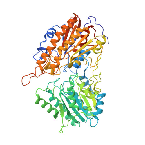
The structures of Leishmania mexicana cofactor-independent phosphoglycerate mutase (Lm iPGAM) crystallised with the substrate 3-phosphoglycerate at high and low cobalt concentrations have been solved at 2.00- and 1.90-A resolutions. Both structures are very similar and the active site contains both 3-phosphoglycerate and 2-phosphoglycerate at equal occupancies (50%). Lm iPGAM co-crystallised with the product 2-phosphoglycerate yields the same structure. Two Co(2+) are coordinated within the active site with different geometries and affinities. The cobalt at the M1 site has a distorted octahedral geometry and is present at 100% occupancy. The M2-site Co(2+) binds with distorted tetrahedral geometry, with only partial occupancy, and coordinates with Ser75, the residue involved in phosphotransfer. When the M2 site is occupied, the side chain of Ser75 adopts a position that is unfavourable for catalysis, indicating that this site may not be occupied under physiological conditions and that catalysis may occur via a one-metal mechanism. The geometry of the M2 site suggests that it is possible for Ser75 to be activated for phosphotransfer by H-bonding to nearby residues rather than by metal coordination. The 16 active-site residues of Lm iPGAM are conserved in the Mn-dependent iPGAM from Bacillus stearothermophilus (33% overall sequence identity). However, Lm iPGAM has an inserted tyrosine (Tyr210) that causes the M2 site to diminish in size, consistent with its reduced metal affinity. Tyr210 is present in trypanosomatid and plant iPGAMs, but not in the enzymes from other organisms, indicating that there are two subclasses of iPGAMs.
Organizational Affiliation:
Structural Biochemistry Group, Institute of Structural and Molecular Biology, University of Edinburgh, King's Buildings, Edinburgh, UK.


