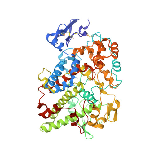
3KK6

Crystal Structure of Cyclooxygenase-1 in complex with celecoxib


Experimental Data Snapshot

  • Method: X-RAY DIFFRACTION
  • Resolution: 2.75 Å
  • R-Value Free: 0.242 
  • R-Value Work: 0.207 
  • R-Value Observed: 0.208 

Starting Model: experimental
View more details

wwPDB Validation   3D Report Full Report


Ligand Structure Quality Assessment 


This is version 2.2 of the entry. See complete history


Literature

Coxibs interfere with the action of aspirin by binding tightly to one monomer of cyclooxygenase-1.

Rimon, G.Sidhu, R.S.Lauver, D.A.Lee, J.Y.Sharma, N.P.Yuan, C.Frieler, R.A.Trievel, R.C.Lucchesi, B.R.Smith, W.L.

(2010) Proc Natl Acad Sci U S A 107: 28-33

  • DOI: https://doi.org/10.1073/pnas.0909765106
  • Primary Citation of Related Structures:  
    3KK6

  • PubMed Abstract: 

    Pain associated with inflammation involves prostaglandins synthesized from arachidonic acid (AA) through cyclooxygenase-2 (COX-2) pathways while thromboxane A(2) formed by platelets from AA via cyclooxygenase-1 (COX-1) mediates thrombosis. COX-1 and COX-2 are both targets of nonselective nonsteroidal antiinflammatory drugs (nsNSAIDs) including aspirin whereas COX-2 activity is preferentially blocked by COX-2 inhibitors called coxibs. COXs are homodimers composed of identical subunits, but we have shown that only one subunit is active at a time during catalysis; moreover, many nsNSAIDS bind to a single subunit of a COX dimer to inhibit the COX activity of the entire dimer. Here, we report the surprising observation that celecoxib and other coxibs bind tightly to a subunit of COX-1. Although celecoxib binding to one monomer of COX-1 does not affect the normal catalytic processing of AA by the second, partner subunit, celecoxib does interfere with the inhibition of COX-1 by aspirin in vitro. X-ray crystallographic results obtained with a celecoxib/COX-1 complex show how celecoxib can bind to one of the two available COX sites of the COX-1 dimer. Finally, we find that administration of celecoxib to dogs interferes with the ability of a low dose of aspirin to inhibit AA-induced ex vivo platelet aggregation. COX-2 inhibitors such as celecoxib are widely used for pain relief. Because coxibs exhibit cardiovascular side effects, they are often prescribed in combination with low-dose aspirin to prevent thrombosis. Our studies predict that the cardioprotective effect of low-dose aspirin on COX-1 may be blunted when taken with coxibs.


  • Organizational Affiliation

    Department of Biological Chemistry, University of Michigan, Ann Arbor, MI 4810, USA.


Macromolecules
Find similar proteins by:  (by identity cutoff)  |  3D Structure
Entity ID: 1
MoleculeChains Sequence LengthOrganismDetailsImage
Prostaglandin G/H synthase 1
A, B
553Ovis ariesMutation(s): 0 
Gene Names: COX1PTGS1
EC: 1.14.99.1
UniProt
Find proteins for P05979 (Ovis aries)
Explore P05979 
Go to UniProtKB:  P05979
Entity Groups  
Sequence Clusters30% Identity50% Identity70% Identity90% Identity95% Identity100% Identity
UniProt GroupP05979
Sequence Annotations
Expand
  • Reference Sequence
Oligosaccharides

Help

Entity ID: 2
MoleculeChains Length2D Diagram Glycosylation3D Interactions
alpha-D-mannopyranose-(1-3)-beta-D-mannopyranose-(1-4)-alpha-D-mannopyranose-(1-4)-2-acetamido-2-deoxy-beta-D-glucopyranose-(1-4)-2-acetamido-2-deoxy-beta-D-glucopyranose
C
5N/A
Glycosylation Resources
GlyTouCan:  G19294PB
GlyCosmos:  G19294PB
GlyGen:  G19294PB
Entity ID: 3
MoleculeChains Length2D Diagram Glycosylation3D Interactions
2-acetamido-2-deoxy-alpha-D-glucopyranose-(1-4)-2-acetamido-2-deoxy-beta-D-glucopyranose
D, E, G
2N/A
Glycosylation Resources
GlyTouCan:  G07375KG
GlyCosmos:  G07375KG
GlyGen:  G07375KG
Entity ID: 4
MoleculeChains Length2D Diagram Glycosylation3D Interactions
beta-D-mannopyranose-(1-6)-alpha-D-mannopyranose-(1-4)-2-acetamido-2-deoxy-beta-D-glucopyranose-(1-4)-2-acetamido-2-deoxy-beta-D-glucopyranose
F
4N/A
Glycosylation Resources
GlyTouCan:  G26644CA
GlyCosmos:  G26644CA
GlyGen:  G26644CA
Small Molecules
Ligands 4 Unique
IDChains Name / Formula / InChI Key2D Diagram3D Interactions
HEM
Query on HEM

Download Ideal Coordinates CCD File 
H [auth A],
M [auth B]
PROTOPORPHYRIN IX CONTAINING FE
C34 H32 Fe N4 O4
KABFMIBPWCXCRK-RGGAHWMASA-L
CEL
Query on CEL

Download Ideal Coordinates CCD File 
I [auth A],
N [auth B]
4-[5-(4-METHYLPHENYL)-3-(TRIFLUOROMETHYL)-1H-PYRAZOL-1-YL]BENZENESULFONAMIDE
C17 H14 F3 N3 O2 S
RZEKVGVHFLEQIL-UHFFFAOYSA-N
BOG
Query on BOG

Download Ideal Coordinates CCD File 
J [auth A],
K [auth A],
O [auth B],
P [auth B]
octyl beta-D-glucopyranoside
C14 H28 O6
HEGSGKPQLMEBJL-RKQHYHRCSA-N
FLC
Query on FLC

Download Ideal Coordinates CCD File 
L [auth A]CITRATE ANION
C6 H5 O7
KRKNYBCHXYNGOX-UHFFFAOYSA-K
Experimental Data & Validation

Experimental Data

  • Method: X-RAY DIFFRACTION
  • Resolution: 2.75 Å
  • R-Value Free: 0.242 
  • R-Value Work: 0.207 
  • R-Value Observed: 0.208 
  • Space Group: P 65
Unit Cell:
Length ( Å )Angle ( ˚ )
a = 181.035α = 90
b = 181.035β = 90
c = 102.698γ = 120
Software Package:
Software NamePurpose
DENZOdata reduction
SCALEPACKdata scaling
PHASERphasing
PHENIXrefinement
PDB_EXTRACTdata extraction

Structure Validation

View Full Validation Report


Ligand Structure Quality Assessment 


Entry History 

Deposition Data

  • Released Date: 2009-12-15 
  • Deposition Author(s): Sidhu, R.S.

Revision History  (Full details and data files)

  • Version 1.0: 2009-12-15
    Type: Initial release
  • Version 1.1: 2011-07-13
    Changes: Non-polymer description, Version format compliance
  • Version 1.2: 2013-03-13
    Changes: Refinement description
  • Version 1.3: 2017-11-01
    Changes: Advisory, Refinement description
  • Version 2.0: 2020-07-29
    Type: Remediation
    Reason: Carbohydrate remediation
    Changes: Advisory, Atomic model, Data collection, Derived calculations, Structure summary
  • Version 2.1: 2023-09-06
    Changes: Advisory, Data collection, Database references, Refinement description, Structure summary
  • Version 2.2: 2024-11-20
    Changes: Structure summary