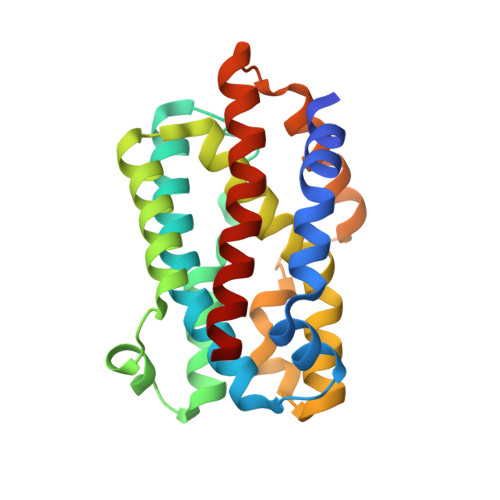
3MOO

Crystal structure of the HmuO, heme oxygenase from Corynebacterium diphtheriae, in complex with azide-bound verdoheme


Experimental Data Snapshot

  • Method: X-RAY DIFFRACTION
  • Resolution: 1.71 Å
  • R-Value Free: 0.213 
  • R-Value Work: 0.178 
  • R-Value Observed: 0.182 

Starting Model: experimental
View more details

wwPDB Validation   3D Report Full Report


Ligand Structure Quality Assessment 


This is version 2.1 of the entry. See complete history


Literature

Enzymatic ring-opening mechanism of verdoheme by the heme oxygenase: a combined X-ray crystallography and QM/MM study.

Lai, W.Chen, H.Matsui, T.Omori, K.Unno, M.Ikeda-Saito, M.Shaik, S.

(2010) J Am Chem Soc 132: 12960-12970

  • DOI: https://doi.org/10.1021/ja104674q
  • Primary Citation of Related Structures:  
    3MOO

  • PubMed Abstract: 

    The least understood mechanism during heme degradation by the enzyme heme oxygenase (HO) is the third step of ring opening of verdoheme to biliverdin, a process which maintains iron homeostasis. In response to this mechanistic uncertainty, we launched a combined study of X-ray crystallography and theoretical QM/MM calculations, designed to elucidate the mechanism. The air-sensitive ferrous verdoheme complex of HmuO, a heme oxygenase from Corynebacterium diphtheriae, was crystallized under anaerobic conditions. Spectral analysis of the azide-bound verdoheme-HmuO complex crystals assures that the verdoheme group remains intact during the crystallization and X-ray diffraction measurement. The structure offers the first solid evidence for the presence of a water cluster in the distal pocket of this catalytically critical intermediate. The subsequent QM/MM calculations based on this crystal structure explore the reaction mechanisms starting from the FeOOH-verdoheme and FeHOOH-verdoheme complexes, which mimic, respectively, the O(2)- and H(2)O(2)-supported degradations. In both mechanisms, the rate-determining step is the initial O-O bond breaking step, which is either homolytic (for FeHOOH-verdoheme) or coupled to electron and proton transfers (in FeOOH-verdoheme). Additionally, the calculations indicate that the FeHOOH-verdoheme complex is more reactive than the FeOOH-verdoheme complex in accord with experimental findings. QM energies with embedded MM charges are close to and yield the same conclusions as full QM/MM energies. Finally, the calculations highlight the dominant influence of the distal water cluster which acts as a biocatalyst for the conversion of verdoheme to biliverdin in the two processes, by fixing the departing OH and directing it to the requisite site of attack, and by acting as a proton shuttle and a haven for the highly reactive OH(-) nucleophile.


  • Organizational Affiliation

    Institute of Chemistry and The Lise Meitner-Minerva Center for Computational Quantum Chemistry, The Hebrew University of Jerusalem, 91904 Jerusalem, Israel.


Macromolecules
Find similar proteins by:  (by identity cutoff)  |  3D Structure
Entity ID: 1
MoleculeChains Sequence LengthOrganismDetailsImage
Heme oxygenase
A, B
215Corynebacterium diphtheriaeMutation(s): 0 
EC: 1.14.14.18
UniProt
Find proteins for Q54AI1 (Corynebacterium diphtheriae)
Explore Q54AI1 
Go to UniProtKB:  Q54AI1
Entity Groups  
Sequence Clusters30% Identity50% Identity70% Identity90% Identity95% Identity100% Identity
UniProt GroupQ54AI1
Sequence Annotations
Expand
  • Reference Sequence
Oligosaccharides

Help

Entity ID: 2
MoleculeChains Length2D Diagram Glycosylation3D Interactions
beta-D-fructofuranose-(2-1)-alpha-D-glucopyranose
C, D
2N/A
Glycosylation Resources
GlyTouCan:  G05551OP
GlyCosmos:  G05551OP
Small Molecules
Ligands 3 Unique
IDChains Name / Formula / InChI Key2D Diagram3D Interactions
VEA
Query on VEA

Download Ideal Coordinates CCD File 
K [auth A],
R [auth B]
5-OXA-PROTOPORPHYRIN IX CONTAINING FE
C33 H31 Fe N4 O5
OCHHJFVQYXRHAA-HPQJSUICSA-M
SO4
Query on SO4

Download Ideal Coordinates CCD File 
F [auth A]
G [auth A]
H [auth A]
I [auth A]
J [auth A]
F [auth A],
G [auth A],
H [auth A],
I [auth A],
J [auth A],
M [auth B],
N [auth B],
O [auth B],
P [auth B],
Q [auth B]
SULFATE ION
O4 S
QAOWNCQODCNURD-UHFFFAOYSA-L
AZI
Query on AZI

Download Ideal Coordinates CCD File 
E [auth A],
L [auth B]
AZIDE ION
N3
IVRMZWNICZWHMI-UHFFFAOYSA-N
Biologically Interesting Molecules (External Reference) 1 Unique
Experimental Data & Validation

Experimental Data

  • Method: X-RAY DIFFRACTION
  • Resolution: 1.71 Å
  • R-Value Free: 0.213 
  • R-Value Work: 0.178 
  • R-Value Observed: 0.182 
  • Space Group: C 1 2 1
Unit Cell:
Length ( Å )Angle ( ˚ )
a = 106.609α = 90
b = 63.649β = 130.34
c = 78.52γ = 90
Software Package:
Software NamePurpose
REFMACrefinement

Structure Validation

View Full Validation Report


Ligand Structure Quality Assessment 


Entry History 

Deposition Data

Revision History  (Full details and data files)

  • Version 1.0: 2011-03-09
    Type: Initial release
  • Version 1.1: 2011-07-13
    Changes: Version format compliance
  • Version 2.0: 2020-07-29
    Type: Remediation
    Reason: Carbohydrate remediation
    Changes: Advisory, Atomic model, Data collection, Derived calculations, Non-polymer description, Structure summary
  • Version 2.1: 2023-11-01
    Changes: Data collection, Database references, Refinement description, Structure summary