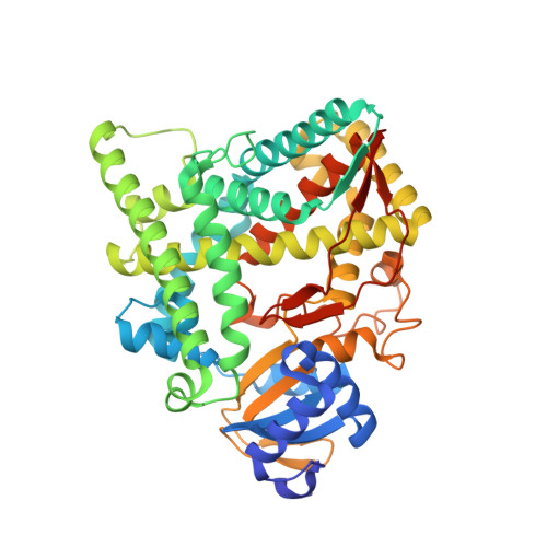
3N9Z

Crystal structure of human CYP11A1 in complex with 22-hydroxycholesterol


Experimental Data Snapshot

  • Method: X-RAY DIFFRACTION
  • Resolution: 2.17 Å
  • R-Value Free: 0.243 
  • R-Value Work: 0.210 
  • R-Value Observed: 0.211 

wwPDB Validation   3D Report Full Report


Ligand Structure Quality Assessment 


This is version 1.3 of the entry. See complete history


Literature

Structural basis for pregnenolone biosynthesis by the mitochondrial monooxygenase system.

Strushkevich, N.Mackenzie, F.Cherkesova, T.Grabovec, I.Usanov, S.Park, H.W.

(2011) Proc Natl Acad Sci U S A 108: 10139-10143

  • DOI: https://doi.org/10.1073/pnas.1019441108
  • Primary Citation of Related Structures:  
    3N9Y, 3N9Z, 3NA0, 3NA1

  • PubMed Abstract: 

    In humans, the precursor to all steroid hormones, pregnenolone, is synthesized from cholesterol by an enzyme complex comprising adrenodoxin reductase (AdR), adrenodoxin (Adx), and a cytochrome P450 (P450scc or CYP11A1). This complex not only plays a key role in steroidogenesis, but also has long been a model to study electron transfer, multistep catalysis, and C-C bond cleavage performed by monooxygenases. Detailed mechanistic understanding of these processes has been hindered by a lack of structural information. Here we present the crystal structure of the complex of human Adx and CYP11A1--the first of a complex between a eukaryotic CYP and its redox partner. The structures with substrate and a series of reaction intermediates allow us to define the mechanism underlying sequential hydroxylations of the cholesterol and suggest the mechanism of C-C bond cleavage. In the complex the [2Fe-2S] cluster of Adx is positioned 17.4 Å away from the heme iron of CYP11A1. This structure suggests that after an initial protein-protein association driven by electrostatic forces, the complex adopts an optimized geometry between the redox centers. Conservation of the interaction interface suggests that this mechanism is common for all mitochondrial P450s.


  • Organizational Affiliation

    Structural Genomics Consortium, University of Toronto, 101 College Street, Toronto, ON, Canada M5G 1L7.


Macromolecules
Find similar proteins by:  (by identity cutoff)  |  3D Structure
Entity ID: 1
MoleculeChains Sequence LengthOrganismDetailsImage
Cholesterol side-chain cleavage enzyme
A, B
487Homo sapiensMutation(s): 0 
Gene Names: CYP11ACYP11A1
EC: 1.14.15.6
UniProt & NIH Common Fund Data Resources
Find proteins for P05108 (Homo sapiens)
Explore P05108 
Go to UniProtKB:  P05108
PHAROS:  P05108
GTEx:  ENSG00000140459 
Entity Groups  
Sequence Clusters30% Identity50% Identity70% Identity90% Identity95% Identity100% Identity
UniProt GroupP05108
Sequence Annotations
Expand
  • Reference Sequence
Find similar proteins by:  (by identity cutoff)  |  3D Structure
Entity ID: 2
MoleculeChains Sequence LengthOrganismDetailsImage
Adrenodoxin
C, D
123Homo sapiensMutation(s): 0 
Gene Names: adrenodoxinADXFDX1
UniProt & NIH Common Fund Data Resources
Find proteins for P10109 (Homo sapiens)
Explore P10109 
Go to UniProtKB:  P10109
PHAROS:  P10109
GTEx:  ENSG00000137714 
Entity Groups  
Sequence Clusters30% Identity50% Identity70% Identity90% Identity95% Identity100% Identity
UniProt GroupP10109
Sequence Annotations
Expand
  • Reference Sequence
Small Molecules
Ligands 3 Unique
IDChains Name / Formula / InChI Key2D Diagram3D Interactions
HEM
Query on HEM

Download Ideal Coordinates CCD File 
E [auth A],
G [auth B]
PROTOPORPHYRIN IX CONTAINING FE
C34 H32 Fe N4 O4
KABFMIBPWCXCRK-RGGAHWMASA-L
HC9
Query on HC9

Download Ideal Coordinates CCD File 
F [auth A],
H [auth B]
(3alpha,8alpha,22R)-cholest-5-ene-3,22-diol
C27 H46 O2
RZPAXNJLEKLXNO-GFKLAVDKSA-N
FES
Query on FES

Download Ideal Coordinates CCD File 
I [auth C],
J [auth D]
FE2/S2 (INORGANIC) CLUSTER
Fe2 S2
NIXDOXVAJZFRNF-UHFFFAOYSA-N
Experimental Data & Validation

Experimental Data

  • Method: X-RAY DIFFRACTION
  • Resolution: 2.17 Å
  • R-Value Free: 0.243 
  • R-Value Work: 0.210 
  • R-Value Observed: 0.211 
  • Space Group: P 1 21 1
Unit Cell:
Length ( Å )Angle ( ˚ )
a = 83.383α = 90
b = 115.095β = 101.82
c = 86.213γ = 90
Software Package:
Software NamePurpose
DENZOdata reduction
SCALEPACKdata scaling
REFMACrefinement
PDB_EXTRACTdata extraction

Structure Validation

View Full Validation Report


Ligand Structure Quality Assessment 


Entry History 

Revision History  (Full details and data files)

  • Version 1.0: 2011-06-08
    Type: Initial release
  • Version 1.1: 2011-07-13
    Changes: Version format compliance
  • Version 1.2: 2017-11-08
    Changes: Refinement description
  • Version 1.3: 2024-02-21
    Changes: Data collection, Database references, Derived calculations