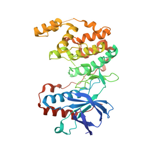
3OEF

Crystal structure of Y323F inactive mutant of p38alpha MAP kinase


Experimental Data Snapshot

  • Method: X-RAY DIFFRACTION
  • Resolution: 1.60 Å
  • R-Value Free: 0.221 
  • R-Value Work: 0.183 
  • R-Value Observed: 0.184 

wwPDB Validation   3D Report Full Report


Ligand Structure Quality Assessment 


This is version 1.4 of the entry. See complete history


Literature

Active mutants of the TCR-mediated p38alpha alternative activation site show changes in the phosphorylation lip and DEF site formation.

Tzarum, N.Diskin, R.Engelberg, D.Livnah, O.

(2011) J Mol Biol 405: 1154-1169

  • DOI: https://doi.org/10.1016/j.jmb.2010.11.023
  • Primary Citation of Related Structures:  
    3OD6, 3ODY, 3ODZ, 3OEF

  • PubMed Abstract: 

    The p38α mitogen-activated protein kinase is commonly activated by dual (Thr and Tyr) phosphorylation catalyzed by mitogen-activated protein kinase kinases. However, in T-cells, upon stimulation of the T-cell receptor, p38α is activated via an alternative pathway, involving its phosphorylation by zeta-chain-associated protein kinase 70 on Tyr323, distal from the phosphorylation lip. Tyr323-phosphorylated p38α is autoactivated, resulting in monophosphorylation of Thr180. The conformational changes induced by pTyr323 mediating autoactivation are not known. The lack of pTyr323 p38α for structural studies promoted the search for Tyr323 mutations that may functionally emulate its effect when phosphorylated. Via a comprehensive mutagenesis of Tyr323, we identified mutations that rendered the kinase intrinsically active and others that displayed no activity. Crystallographic studies of selected active (p38α(Y323Q), p38α(Y323T), and p38α(Y323R)) and inactive (p38α(Y323F)) mutants revealed that substantial changes in interlobe orientation, extended conformation of the activation loop, and formation of substrate docking DEF site (docking site for extracellular signal-regulated kinase FXF) interaction pocket are associated with p38α activation.


  • Organizational Affiliation

    Department of Biological Chemistry, Alexander Silberman Institute of Life Sciences, Hebrew University of Jerusalem, Jerusalem 91904, Israel.


Macromolecules
Find similar proteins by:  (by identity cutoff)  |  3D Structure
Entity ID: 1
MoleculeChains Sequence LengthOrganismDetailsImage
Mitogen-activated protein kinase 14A [auth X]360Homo sapiensMutation(s): 1 
Gene Names: MAPK14CSBPCSBP1CSBP2CSPB1MXI2
EC: 2.7.11.24
UniProt & NIH Common Fund Data Resources
Find proteins for Q16539 (Homo sapiens)
Explore Q16539 
Go to UniProtKB:  Q16539
PHAROS:  Q16539
GTEx:  ENSG00000112062 
Entity Groups  
Sequence Clusters30% Identity50% Identity70% Identity90% Identity95% Identity100% Identity
UniProt GroupQ16539
Sequence Annotations
Expand
  • Reference Sequence
Experimental Data & Validation

Experimental Data

  • Method: X-RAY DIFFRACTION
  • Resolution: 1.60 Å
  • R-Value Free: 0.221 
  • R-Value Work: 0.183 
  • R-Value Observed: 0.184 
  • Space Group: P 21 21 21
Unit Cell:
Length ( Å )Angle ( ˚ )
a = 64.734α = 90
b = 69.07β = 90
c = 74.409γ = 90
Software Package:
Software NamePurpose
HKL-2000data collection
MOLREPphasing
PHENIXrefinement
HKL-2000data reduction
HKL-2000data scaling

Structure Validation

View Full Validation Report


Ligand Structure Quality Assessment 


Entry History 

Deposition Data

Revision History  (Full details and data files)

  • Version 1.0: 2011-01-12
    Type: Initial release
  • Version 1.1: 2011-07-13
    Changes: Version format compliance
  • Version 1.2: 2013-07-03
    Changes: Database references
  • Version 1.3: 2020-07-29
    Type: Remediation
    Reason: Carbohydrate remediation
    Changes: Data collection, Database references, Derived calculations, Structure summary
  • Version 1.4: 2024-02-21
    Changes: Data collection, Database references, Structure summary