3PDB

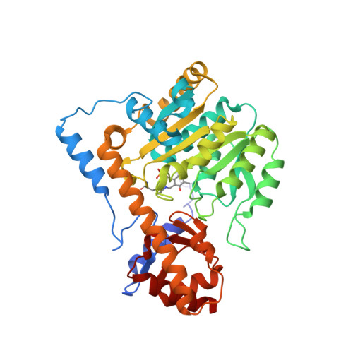
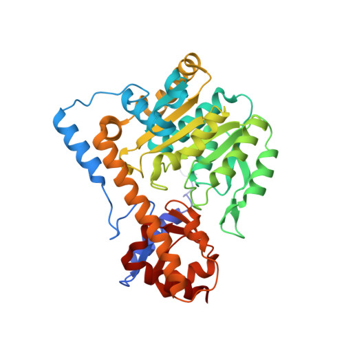
Crystal structure of mouse mitochondrial aspartate aminotransferase in complex with oxaloacetic acid


Experimental Data Snapshot

  • Method: X-RAY DIFFRACTION
  • Resolution: 2.40 Å
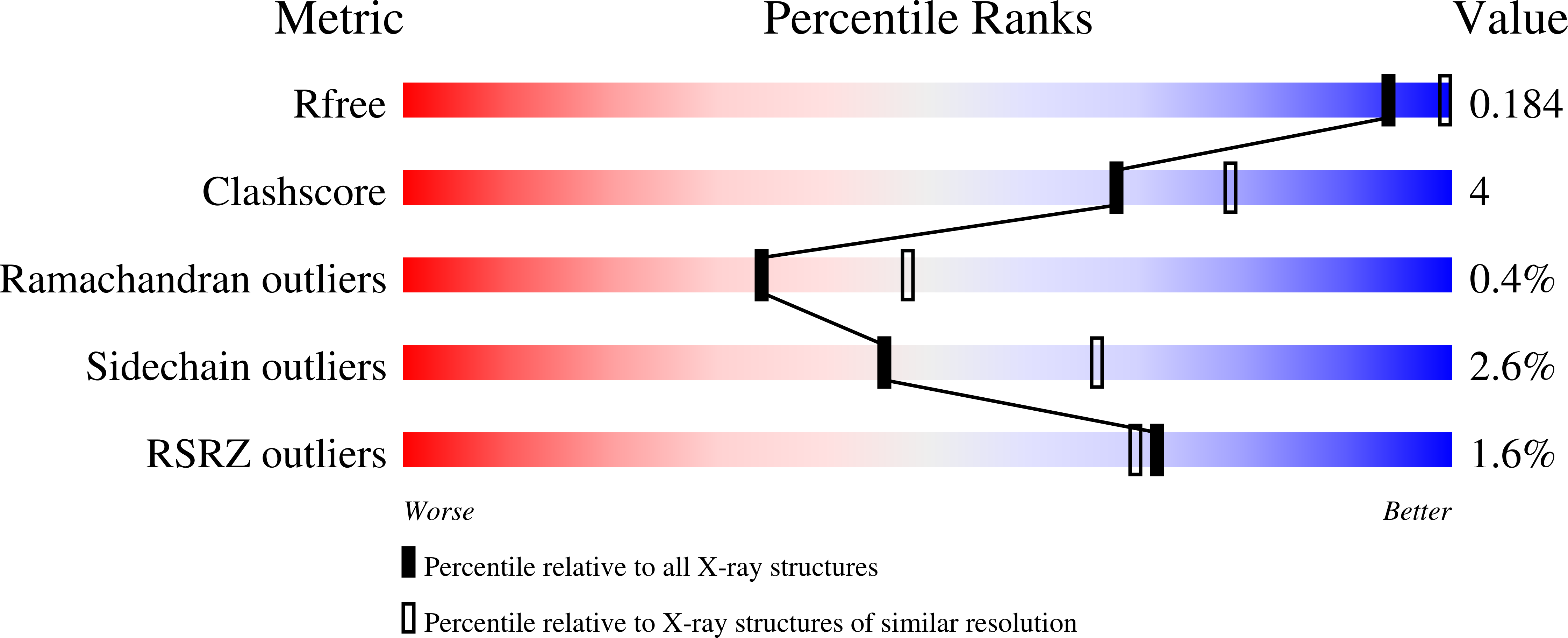
  • R-Value Free: 0.190 
  • R-Value Work: 0.177 
  • R-Value Observed: 0.178 

Starting Model: experimental
View more details

wwPDB Validation   3D Report Full Report


Ligand Structure Quality Assessment 


This is version 1.3 of the entry. See complete history


Literature

Biochemical and structural characterization of mouse mitochondrial aspartate aminotransferase, a newly identified kynurenine aminotransferase-IV.

Han, Q.Robinson, H.Cai, T.Tagle, D.A.Li, J.

(2011) Biosci Rep 31: 323-332

  • DOI: https://doi.org/10.1042/BSR20100117
  • Primary Citation of Related Structures:  
    3PD6, 3PDB

  • PubMed Abstract: 

    Mammalian mAspAT (mitochondrial aspartate aminotransferase) is recently reported to have KAT (kynurenine aminotransferase) activity and plays a role in the biosynthesis of KYNA (kynurenic acid) in rat, mouse and human brains. This study concerns the biochemical and structural characterization of mouse mAspAT. In this study, mouse mAspAT cDNA was amplified from mouse brain first stand cDNA and its recombinant protein was expressed in an Escherichia coli expression system. Sixteen oxo acids were tested for the co-substrate specificity of mouse mAspAT and 14 of them were shown to be capable of serving as co-substrates for the enzyme. Structural analysis of mAspAT by macromolecular crystallography revealed that the cofactor-binding residues of mAspAT are similar to those of other KATs. The substrate-binding residues of mAspAT are slightly different from those of other KATs. Our results provide a biochemical and structural basis towards understanding the overall physiological role of mAspAT in vivo and insight into controlling the levels of endogenous KYNA through modulation of the enzyme in the mouse brain.


  • Organizational Affiliation

    Department of Biochemistry, Virginia Tech, Blacksburg, VA 24061, USA.


Macromolecules
Find similar proteins by:  (by identity cutoff)  |  3D Structure
Entity ID: 1
MoleculeChains Sequence LengthOrganismDetailsImage
Aspartate aminotransferase, mitochondrial
A, C
401Mus musculusMutation(s): 0 
Gene Names: GOT2
EC: 2.6.1.1 (PDB Primary Data), 2.6.1.7 (UniProt)
UniProt & NIH Common Fund Data Resources
Find proteins for P05202 (Mus musculus)
Explore P05202 
Go to UniProtKB:  P05202
IMPC:  MGI:95792
Entity Groups  
Sequence Clusters30% Identity50% Identity70% Identity90% Identity95% Identity100% Identity
UniProt GroupP05202
Sequence Annotations
Expand
  • Reference Sequence
Find similar proteins by:  (by identity cutoff)  |  3D Structure
Entity ID: 2
MoleculeChains Sequence LengthOrganismDetailsImage
Aspartate aminotransferase, mitochondrial
B, D
401Mus musculusMutation(s): 0 
Gene Names: GOT2
EC: 2.6.1.1 (PDB Primary Data), 2.6.1.7 (UniProt)
UniProt & NIH Common Fund Data Resources
Find proteins for P05202 (Mus musculus)
Explore P05202 
Go to UniProtKB:  P05202
IMPC:  MGI:95792
Entity Groups  
Sequence Clusters30% Identity50% Identity70% Identity90% Identity95% Identity100% Identity
UniProt GroupP05202
Sequence Annotations
Expand
  • Reference Sequence
Small Molecules
Ligands 4 Unique
IDChains Name / Formula / InChI Key2D Diagram3D Interactions
PMP
Query on PMP

Download Ideal Coordinates CCD File 
J [auth B],
Q [auth D]
4'-DEOXY-4'-AMINOPYRIDOXAL-5'-PHOSPHATE
C8 H13 N2 O5 P
ZMJGSOSNSPKHNH-UHFFFAOYSA-N
OAA
Query on OAA

Download Ideal Coordinates CCD File 
H [auth B]OXALOACETATE ION
C4 H3 O5
KHPXUQMNIQBQEV-UHFFFAOYSA-M
GOL
Query on GOL

Download Ideal Coordinates CCD File 
F [auth A],
G [auth A],
N [auth C],
R [auth D],
S [auth D]
GLYCEROL
C3 H8 O3
PEDCQBHIVMGVHV-UHFFFAOYSA-N
BME
Query on BME

Download Ideal Coordinates CCD File 
E [auth A]
I [auth B]
K [auth B]
L [auth C]
M [auth C]
E [auth A],
I [auth B],
K [auth B],
L [auth C],
M [auth C],
O [auth D],
P [auth D]
BETA-MERCAPTOETHANOL
C2 H6 O S
DGVVWUTYPXICAM-UHFFFAOYSA-N
Modified Residues  1 Unique
IDChains TypeFormula2D DiagramParent
LLP
Query on LLP
A, C
L-PEPTIDE LINKINGC14 H22 N3 O7 PLYS
Experimental Data & Validation

Experimental Data

  • Method: X-RAY DIFFRACTION
  • Resolution: 2.40 Å
  • R-Value Free: 0.190 
  • R-Value Work: 0.177 
  • R-Value Observed: 0.178 
  • Space Group: P 21 21 2
Unit Cell:
Length ( Å )Angle ( ˚ )
a = 282.418α = 90
b = 77.894β = 90
c = 87.389γ = 90
Software Package:
Software NamePurpose
HKL-2000data collection
MOLREPphasing
REFMACrefinement
HKL-2000data reduction
HKL-2000data scaling

Structure Validation

View Full Validation Report


Ligand Structure Quality Assessment 


Entry History 

Deposition Data

Revision History  (Full details and data files)

  • Version 1.0: 2010-11-10
    Type: Initial release
  • Version 1.1: 2011-07-13
    Changes: Version format compliance
  • Version 1.2: 2023-09-06
    Changes: Data collection, Database references, Derived calculations, Refinement description
  • Version 1.3: 2023-12-06
    Changes: Data collection