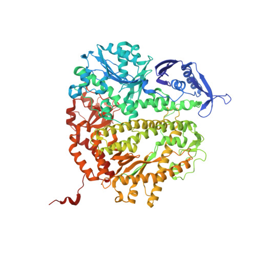
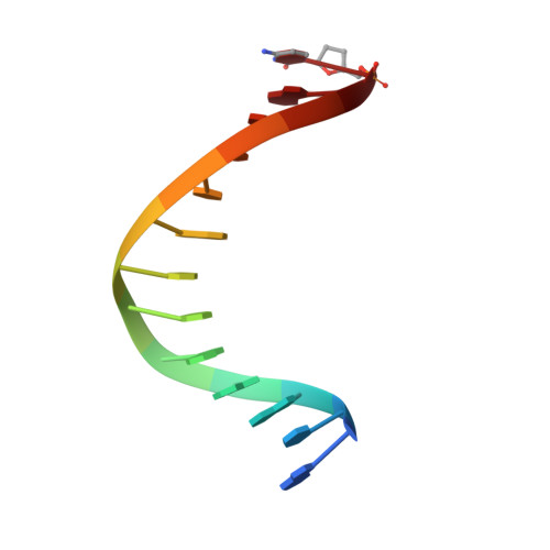
3S9H

RB69 DNA Polymerase Triple Mutant(L561A/S565G/Y567A) ternary complex with dUpNpp and a dideoxy-terminated primer in the presence of Ca2+


Experimental Data Snapshot

  • Method: X-RAY DIFFRACTION
  • Resolution: 1.95 Å
  • R-Value Free: 0.213 
  • R-Value Work: 0.171 
  • R-Value Observed: 0.173 

Starting Model: experimental
View more details

wwPDB Validation   3D Report Full Report


Ligand Structure Quality Assessment 


This is version 1.2 of the entry. See complete history


Literature

Structural Insights into Complete Metal Ion Coordination from Ternary Complexes of B Family RB69 DNA Polymerase.

Xia, S.Wang, M.Blaha, G.Konigsberg, W.H.Wang, J.

(2011) Biochemistry 50: 9114-9124

  • DOI: https://doi.org/10.1021/bi201260h
  • Primary Citation of Related Structures:  
    3S9H, 3SCX, 3SI6, 3SJJ, 3SNN, 3SPY, 3SPZ, 3SQ0, 3SQ1

  • PubMed Abstract: 

    We have captured a preinsertion ternary complex of RB69 DNA polymerase (RB69pol) containing the 3' hydroxyl group at the terminus of an extendable primer (ptO3') and a nonhydrolyzable 2'-deoxyuridine 5'-α,β-substituted triphosphate, dUpXpp, where X is either NH or CH(2), opposite a complementary templating dA nucleotide residue. Here we report four structures of these complexes formed by three different RB69pol variants with catalytically inert Ca(2+) and four other structures with catalytically competent Mn(2+) or Mg(2+). These structures provide new insights into why the complete divalent metal-ion coordination complexes at the A and B sites are required for nucleotidyl transfer. They show that the metal ion in the A site brings ptO3' close to the α-phosphorus atom (Pα) of the incoming dNTP to enable phosphodiester bond formation through simultaneous coordination of both ptO3' and the nonbridging Sp oxygen of the dNTP's α-phosphate. The coordination bond length of metal ion A as well as its ionic radius determines how close ptO3' can approach Pα. These variables are expected to affect the rate of bond formation. The metal ion in the B site brings the pyrophosphate product close enough to Pα to enable pyrophosphorolysis and assist in the departure of the pyrophosphate. In these dUpXpp-containing complexes, ptO3' occupies the vertex of a distorted metal ion A coordination octahedron. When ptO3' is placed at the vertex of an undistorted, idealized metal ion A octahedron, it is within bond formation distance to Pα. This geometric relationship appears to be conserved among DNA polymerases of known structure.


  • Organizational Affiliation

    Department of Molecular Biophysics and Biochemistry, Yale University, New Haven, Connecticut 96520-8114, United States.


Macromolecules

Find similar proteins by:  (by identity cutoff)  |  3D Structure
Entity ID: 3
MoleculeChains Sequence LengthOrganismDetailsImage
DNA polymeraseC [auth A]903Escherichia phage RB69Mutation(s): 5 
Gene Names: 43gp43
EC: 2.7.7.7 (PDB Primary Data), 3.1.11 (UniProt)
UniProt
Find proteins for Q38087 (Escherichia phage RB69)
Explore Q38087 
Go to UniProtKB:  Q38087
Entity Groups  
Sequence Clusters30% Identity50% Identity70% Identity90% Identity95% Identity100% Identity
UniProt GroupQ38087
Sequence Annotations
Expand
  • Reference Sequence

Find similar nucleic acids by:  Sequence   |   3D Structure  

Entity ID: 1
MoleculeChains LengthOrganismImage
5'-D(*TP*CP*AP*AP*GP*TP*AP*AP*GP*CP*AP*GP*TP*CP*CP*GP*CP*G)-3'A [auth T]18N/A
Sequence Annotations
Expand
  • Reference Sequence

Find similar nucleic acids by:  Sequence   |   3D Structure  

Entity ID: 2
MoleculeChains LengthOrganismImage
5'-D(*GP*CP*GP*GP*AP*CP*TP*GP*CP*TP*TP*AP*(DOC))-3'B [auth P]13N/A
Sequence Annotations
Expand
  • Reference Sequence
Small Molecules
Ligands 2 Unique
IDChains Name / Formula / InChI Key2D Diagram3D Interactions
DUP
Query on DUP

Download Ideal Coordinates CCD File 
E [auth A]2'-DEOXYURIDINE 5'-ALPHA,BETA-IMIDO-TRIPHOSPHATE
C9 H16 N3 O13 P3
XZLLMTSKYYYJLH-SHYZEUOFSA-N
CA
Query on CA

Download Ideal Coordinates CCD File 
D [auth T]
F [auth A]
G [auth A]
H [auth A]
I [auth A]
D [auth T],
F [auth A],
G [auth A],
H [auth A],
I [auth A],
J [auth A],
K [auth A]
CALCIUM ION
Ca
BHPQYMZQTOCNFJ-UHFFFAOYSA-N
Experimental Data & Validation

Experimental Data

  • Method: X-RAY DIFFRACTION
  • Resolution: 1.95 Å
  • R-Value Free: 0.213 
  • R-Value Work: 0.171 
  • R-Value Observed: 0.173 
  • Space Group: P 21 21 21
Unit Cell:
Length ( Å )Angle ( ˚ )
a = 75.05α = 90
b = 119.821β = 90
c = 130.791γ = 90
Software Package:
Software NamePurpose
HKL-2000data collection
AMoREphasing
REFMACrefinement
HKL-2000data reduction
HKL-2000data scaling

Structure Validation

View Full Validation Report


Ligand Structure Quality Assessment 


Entry History 

Deposition Data

Revision History  (Full details and data files)

  • Version 1.0: 2011-10-12
    Type: Initial release
  • Version 1.1: 2011-11-02
    Changes: Database references
  • Version 1.2: 2023-09-13
    Changes: Data collection, Database references, Derived calculations, Refinement description