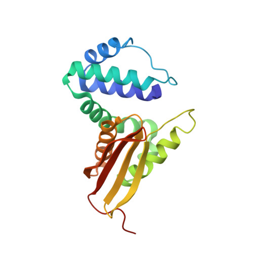
Controlling Conformational Flexibility of an O2-Binding H-NOX Domain.
Weinert, E.E., Phillips-Piro, C.M., Tran, R., Mathies, R.A., Marletta, M.A.(2011) Biochemistry 50: 6832-6840
- PubMed: 21721586
- DOI: https://doi.org/10.1021/bi200788x
- Primary Citation of Related Structures:
3SJ5 - PubMed Abstract:
Heme Nitric oxide/OXygen binding (H-NOX) domains have provided a novel scaffold to probe ligand affinity in hemoproteins. Mutation of isoleucine 5, a conserved residue located in the heme-binding pocket of the H-NOX domain from Thermoanaerobacter tengcongensis (Tt H-NOX), was carried out to examine changes in oxygen (O(2))-binding properties. A series of I5 mutants (I5F, I5F/I75F, I5F/L144F, I5F/I75F/L144F) were investigated to probe the role of steric bulk within the heme pocket. The mutations significantly increased O(2) association rates (1.5-2.5-fold) and dissociation rates (8-190-fold) as compared to wild-type Tt H-NOX. Structural changes that accompanied the I5F mutation were characterized using X-ray crystallography and resonance Raman spectroscopy. A 1.67 Å crystal structure of the I5F mutant indicated that introducing a phenylalanine at position 5 resulted in a significant shift of the N-terminal domain of the protein, causing an opening of the heme pocket. This movement also resulted in an increased amount of flexibility at the N-terminus and the loop covering the N-terminal helix as indicated by the two conformations of the first six N-terminal amino acids, high B-factors in this region of the protein, and partially discontinuous electron density. In addition, introduction of a phenylalanine at position 5 resulted in increased flexibility of the heme within the pocket and weakened hydrogen bonding to the bound O(2) as measured by resonance Raman spectroscopy. This study provides insight into the critical role of I5 in controlling conformational flexibility and ligand affinity in H-NOX proteins.
Organizational Affiliation:
Department of Chemistry, University of California, Berkeley, Berkeley, California 94720, United States.


