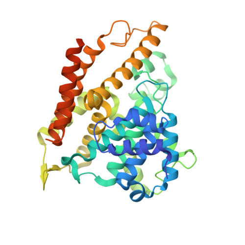
3SL6

Crystal structure of the catalytic domain of PDE4D2 with compound 12c


Experimental Data Snapshot

  • Method: X-RAY DIFFRACTION
  • Resolution: 2.44 Å
  • R-Value Free: 0.256 
  • R-Value Work: 0.198 
  • R-Value Observed: 0.201 

wwPDB Validation   3D Report Full Report


Ligand Structure Quality Assessment 


This is version 1.2 of the entry. See complete history


Literature

Thiophene inhibitors of PDE4: Crystal structures show a second binding mode at the catalytic domain of PDE4D2.

Nankervis, J.L.Feil, S.C.Hancock, N.C.Zheng, Z.Ng, H.L.Morton, C.J.Holien, J.K.Ho, P.W.Frazzetto, M.M.Jennings, I.G.Manallack, D.T.Martin, T.J.Thompson, P.E.Parker, M.W.

(2011) Bioorg Med Chem Lett 21: 7089-7093

  • DOI: https://doi.org/10.1016/j.bmcl.2011.09.109
  • Primary Citation of Related Structures:  
    3SL3, 3SL4, 3SL5, 3SL6, 3SL8

  • PubMed Abstract: 

    PDE4 inhibitors have been identified as therapeutic targets for a variety of conditions, particularly inflammatory diseases. We have serendipitously identified a novel class of phosphodiesterase 4 (PDE4) inhibitor during a study to discover antagonists of the parathyroid hormone receptor. X-ray crystallographic studies of PDE4D2 complexed to four potent inhibitors reveal the atomic details of how they inhibit the enzyme and a notable contrast to another recently reported thiophene-based inhibitor.


  • Organizational Affiliation

    Medicinal Chemistry & Drug Action, Monash Institute of Pharmaceutical Sciences, Monash University (Parkville campus), Parkville, Victoria, Australia.


Macromolecules
Find similar proteins by:  (by identity cutoff)  |  3D Structure
Entity ID: 1
MoleculeChains Sequence LengthOrganismDetailsImage
cAMP-specific 3',5'-cyclic phosphodiesterase 4D
A, B, C, D
361Homo sapiensMutation(s): 0 
Gene Names: PDE4DDPDE3
EC: 3.1.4.17 (PDB Primary Data), 3.1.4.53 (UniProt)
UniProt & NIH Common Fund Data Resources
Find proteins for Q08499 (Homo sapiens)
Explore Q08499 
Go to UniProtKB:  Q08499
PHAROS:  Q08499
GTEx:  ENSG00000113448 
Entity Groups  
Sequence Clusters30% Identity50% Identity70% Identity90% Identity95% Identity100% Identity
UniProt GroupQ08499
Sequence Annotations
Expand
  • Reference Sequence
Small Molecules
Ligands 5 Unique
IDChains Name / Formula / InChI Key2D Diagram3D Interactions
JN8
Query on JN8

Download Ideal Coordinates CCD File 
HA [auth C],
N [auth A],
V [auth B],
VA [auth D]
cyclopentyl 6-(ethylcarbamoyl)-2-[(thiophen-2-ylacetyl)amino]-4,5,6,7-tetrahydrothieno[2,3-c]pyridine-3-carboxylate
C22 H27 N3 O4 S2
TWWIEHZKMVMPFQ-UHFFFAOYSA-N
EPE
Query on EPE

Download Ideal Coordinates CCD File 
AA [auth C]4-(2-HYDROXYETHYL)-1-PIPERAZINE ETHANESULFONIC ACID
C8 H18 N2 O4 S
JKMHFZQWWAIEOD-UHFFFAOYSA-N
DMS
Query on DMS

Download Ideal Coordinates CCD File 
W [auth B]DIMETHYL SULFOXIDE
C2 H6 O S
IAZDPXIOMUYVGZ-UHFFFAOYSA-N
ZN
Query on ZN

Download Ideal Coordinates CCD File 
E [auth A]
F [auth A]
JA [auth D]
KA [auth D]
Q [auth B]
E [auth A],
F [auth A],
JA [auth D],
KA [auth D],
Q [auth B],
R [auth B],
Y [auth C],
Z [auth C]
ZINC ION
Zn
PTFCDOFLOPIGGS-UHFFFAOYSA-N
EDO
Query on EDO

Download Ideal Coordinates CCD File 
BA [auth C]
CA [auth C]
DA [auth C]
EA [auth C]
FA [auth C]
BA [auth C],
CA [auth C],
DA [auth C],
EA [auth C],
FA [auth C],
G [auth A],
GA [auth C],
H [auth A],
I [auth A],
IA [auth D],
J [auth A],
K [auth A],
L [auth A],
LA [auth D],
M [auth A],
MA [auth D],
NA [auth D],
O [auth A],
OA [auth D],
P [auth A],
PA [auth D],
QA [auth D],
RA [auth D],
S [auth B],
SA [auth D],
T [auth B],
TA [auth D],
U [auth B],
UA [auth D],
X [auth C]
1,2-ETHANEDIOL
C2 H6 O2
LYCAIKOWRPUZTN-UHFFFAOYSA-N
Experimental Data & Validation

Experimental Data

  • Method: X-RAY DIFFRACTION
  • Resolution: 2.44 Å
  • R-Value Free: 0.256 
  • R-Value Work: 0.198 
  • R-Value Observed: 0.201 
  • Space Group: P 21 21 21
Unit Cell:
Length ( Å )Angle ( ˚ )
a = 99.54α = 90
b = 111.2β = 90
c = 160.95γ = 90
Software Package:
Software NamePurpose
d*TREKdata scaling
d*TREKdata reduction
PHENIXrefinement
PDB_EXTRACTdata extraction
CrystalCleardata collection
MOLREPphasing

Structure Validation

View Full Validation Report


Ligand Structure Quality Assessment 


Entry History 

Deposition Data

  • Released Date: 2011-10-26 
  • Deposition Author(s): Feil, S.F.

Revision History  (Full details and data files)

  • Version 1.0: 2011-10-26
    Type: Initial release
  • Version 1.1: 2011-12-14
    Changes: Database references
  • Version 1.2: 2024-02-28
    Changes: Data collection, Database references, Derived calculations