3SPG

Inward rectifier potassium channel Kir2.2 R186A mutant in complex with PIP2


Experimental Data Snapshot

  • Method: X-RAY DIFFRACTION
  • Resolution: 2.61 Å
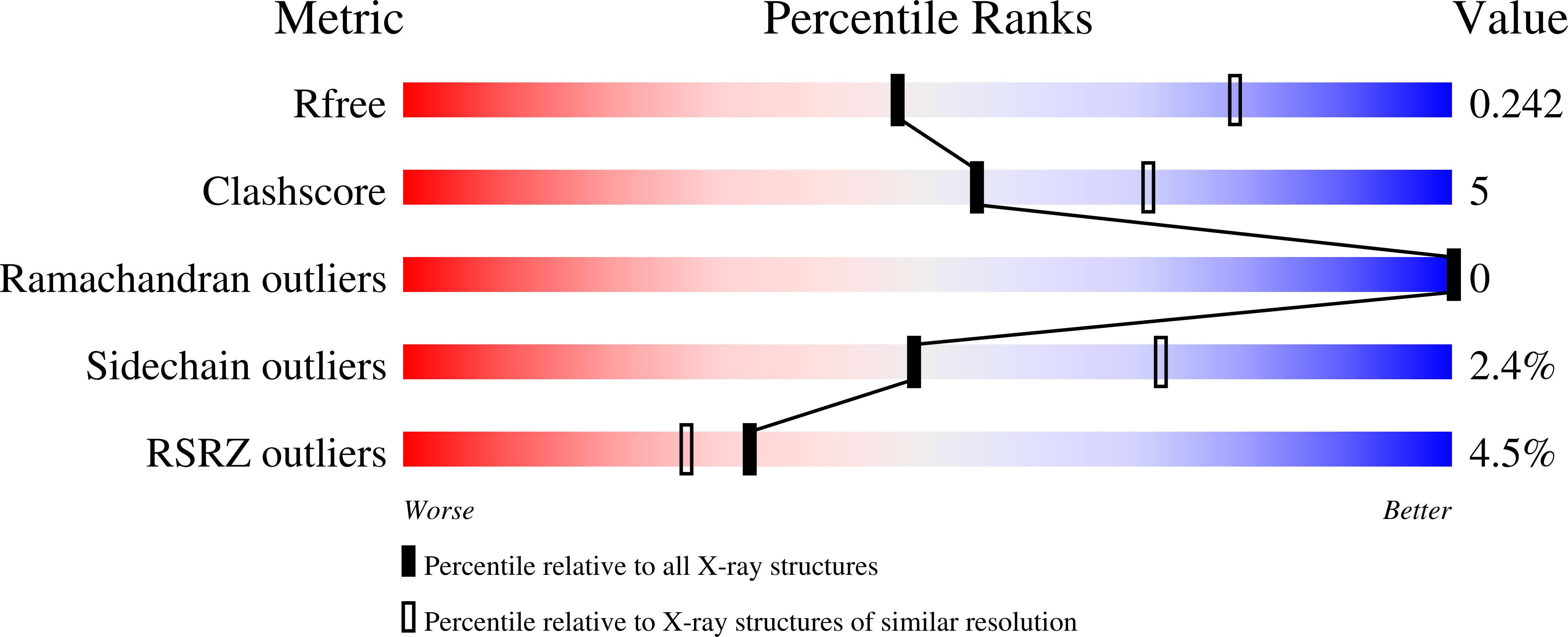
  • R-Value Free: 0.241 
  • R-Value Work: 0.195 
  • R-Value Observed: 0.197 

Starting Model: experimental
View more details

wwPDB Validation   3D Report Full Report


Ligand Structure Quality Assessment 


This is version 1.2 of the entry. See complete history


Literature

Structural basis of PIP(2) activation of the classical inward rectifier K(+) channel Kir2.2.

Hansen, S.B.Tao, X.Mackinnon, R.

(2011) Nature 477: 495-498

  • DOI: https://doi.org/10.1038/nature10370
  • Primary Citation of Related Structures:  
    3SPC, 3SPG, 3SPH, 3SPI, 3SPJ

  • PubMed Abstract: 

    The regulation of ion channel activity by specific lipid molecules is widely recognized as an integral component of electrical signalling in cells. In particular, phosphatidylinositol 4,5-bisphosphate (PIP(2)), a minor yet dynamic phospholipid component of cell membranes, is known to regulate many different ion channels. PIP(2) is the primary agonist for classical inward rectifier (Kir2) channels, through which this lipid can regulate a cell's resting membrane potential. However, the molecular mechanism by which PIP(2) exerts its action is unknown. Here we present the X-ray crystal structure of a Kir2.2 channel in complex with a short-chain (dioctanoyl) derivative of PIP(2). We found that PIP(2) binds at an interface between the transmembrane domain (TMD) and the cytoplasmic domain (CTD). The PIP(2)-binding site consists of a conserved non-specific phospholipid-binding region in the TMD and a specific phosphatidylinositol-binding region in the CTD. On PIP(2) binding, a flexible expansion linker contracts to a compact helical structure, the CTD translates 6 Å and becomes tethered to the TMD and the inner helix gate begins to open. In contrast, the small anionic lipid dioctanoyl glycerol pyrophosphatidic acid (PPA) also binds to the non-specific TMD region, but not to the specific phosphatidylinositol region, and thus fails to engage the CTD or open the channel. Our results show how PIP(2) can control the resting membrane potential through a specific ion-channel-receptor-ligand interaction that brings about a large conformational change, analogous to neurotransmitter activation of ion channels at synapses.


  • Organizational Affiliation

    Laboratory of Molecular Neurobiology & Biophysics, The Rockefeller University, Howard Hughes Medical Institute, 1230 York Avenue, New York, New York 10065, USA.


Macromolecules
Find similar proteins by:  (by identity cutoff)  |  3D Structure
Entity ID: 1
MoleculeChains Sequence LengthOrganismDetailsImage
Inward-rectifier K+ channel Kir2.2343Gallus gallusMutation(s): 1 
Gene Names: Kir2.2
Membrane Entity: Yes 
UniProt
Find proteins for F1NHE9 (Gallus gallus)
Explore F1NHE9 
Go to UniProtKB:  F1NHE9
Entity Groups  
Sequence Clusters30% Identity50% Identity70% Identity90% Identity95% Identity100% Identity
UniProt GroupF1NHE9
Sequence Annotations
Expand
  • Reference Sequence
Small Molecules
Ligands 2 Unique
IDChains Name / Formula / InChI Key2D Diagram3D Interactions
PIO
Query on PIO

Download Ideal Coordinates CCD File 
H [auth A][(2R)-2-octanoyloxy-3-[oxidanyl-[(1R,2R,3S,4R,5R,6S)-2,3,6-tris(oxidanyl)-4,5-diphosphonooxy-cyclohexyl]oxy-phosphoryl]oxy-propyl] octanoate
C25 H49 O19 P3
XLNCEHRXXWQMPK-MJUMVPIBSA-N
K
Query on K

Download Ideal Coordinates CCD File 
B [auth A]
C [auth A]
D [auth A]
E [auth A]
F [auth A]
B [auth A],
C [auth A],
D [auth A],
E [auth A],
F [auth A],
G [auth A]
POTASSIUM ION
K
NPYPAHLBTDXSSS-UHFFFAOYSA-N
Experimental Data & Validation

Experimental Data

  • Method: X-RAY DIFFRACTION
  • Resolution: 2.61 Å
  • R-Value Free: 0.241 
  • R-Value Work: 0.195 
  • R-Value Observed: 0.197 
  • Space Group: I 4
Unit Cell:
Length ( Å )Angle ( ˚ )
a = 81.865α = 90
b = 81.865β = 90
c = 183.102γ = 90
Software Package:
Software NamePurpose
CBASSdata collection
PHENIXrefinement
HKL-2000data reduction
HKL-2000data scaling

Structure Validation

View Full Validation Report


Ligand Structure Quality Assessment 


Entry History 

Deposition Data

Revision History  (Full details and data files)

  • Version 1.0: 2011-08-24
    Type: Initial release
  • Version 1.1: 2011-09-21
    Changes: Database references
  • Version 1.2: 2023-09-13
    Changes: Data collection, Database references, Derived calculations, Refinement description