Crystal Structure of p44, a Constitutively Active Splice Variant of Visual Arrestin.
Granzin, J., Cousin, A., Weirauch, M., Schlesinger, R., Buldt, G., Batra-Safferling, R.(2012) J Mol Biol 416: 611-618
- PubMed: 22306737
- DOI: https://doi.org/10.1016/j.jmb.2012.01.028
- Primary Citation of Related Structures:
3UGU, 3UGX - PubMed Abstract:
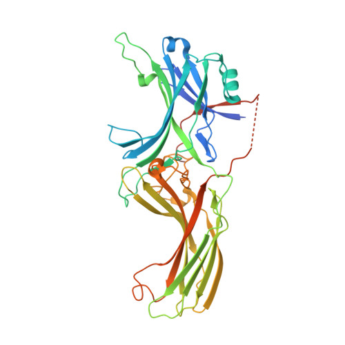
Visual arrestin specifically binds to photoactivated and phosphorylated rhodopsin and inactivates phototransduction. In contrast, the p44 splice variant can terminate phototransduction by binding to nonphosphorylated light-activated rhodopsin. Here we report the crystal structure of bovine p44 at a resolution of 1.85 Å. Compared to native arrestin, the p44 structure reveals significant differences in regions crucial for receptor binding, namely flexible loop V-VI and polar core regions. Additionally, electrostatic potential is remarkably positive on the N-domain and the C-domain. The p44 structure represents an active conformation that serves as a model to explain the 'constitutive activity' found in arrestin variants.
Organizational Affiliation:
Institute of Complex Systems, ICS-6: Structural Biochemistry, Forschungszentrum Jülich, 52425 Jülich, Germany.


