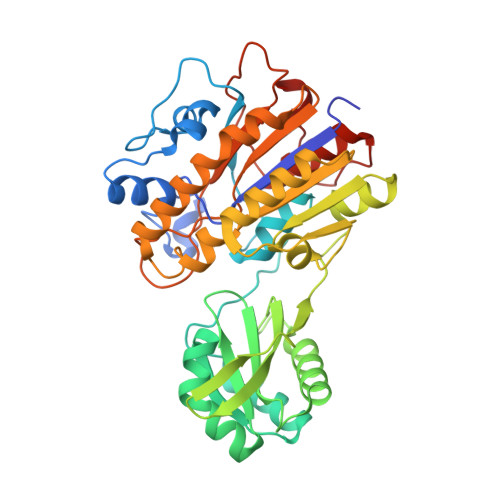
Molecular Differences between a Mutase and a Phosphatase: Investigations of the Activation Step in Bacillus cereus Phosphopentomutase.
Iverson, T.M., Panosian, T.D., Birmingham, W.R., Nannemann, D.P., Bachmann, B.O.(2012) Biochemistry 51: 1964-1975
- PubMed: 22329805
- DOI: https://doi.org/10.1021/bi201761h
- Primary Citation of Related Structures:
3TWZ, 3TX0, 3UN2, 3UN3, 3UN5, 3UNY, 3UO0 - PubMed Abstract:
Prokaryotic phosphopentomutases (PPMs) are di-Mn(2+) enzymes that catalyze the interconversion of α-D-ribose 5-phosphate and α-D-ribose 1-phosphate at an active site located between two independently folded domains. These prokaryotic PPMs belong to the alkaline phosphatase superfamily, but previous studies of Bacillus cereus PPM suggested adaptations of the conserved alkaline phosphatase catalytic cycle. Notably, B. cereus PPM engages substrates when the active site nucleophile, Thr-85, is phosphorylated. Further, the phosphoenzyme is stable throughout purification and crystallization. In contrast, alkaline phosphatase engages substrates when the active site nucleophile is dephosphorylated, and the phosphoenzyme reaction intermediate is only stably trapped in a catalytically compromised enzyme. Studies were undertaken to understand the divergence of these mechanisms. Crystallographic and biochemical investigations of the PPM(T85E) phosphomimetic variant and the neutral corollary PPM(T85Q) determined that the side chain of Lys-240 underwent a change in conformation in response to active site charge, which modestly influenced the affinity for the small molecule activator α-D-glucose 1,6-bisphosphate. More strikingly, the structure of unphosphorylated B. cereus PPM revealed a dramatic change in the interdomain angle and a new hydrogen bonding interaction between the side chain of Asp-156 and the active site nucleophile, Thr-85. This hydrogen bonding interaction is predicted to align and activate Thr-85 for nucleophilic addition to α-D-glucose 1,6-bisphosphate, favoring the observed equilibrium phosphorylated state. Indeed, phosphorylation of Thr-85 is severely impaired in the PPM(D156A) variant even under stringent activation conditions. These results permit a proposal for activation of PPM and explain some of the essential features that distinguish between the catalytic cycles of PPM and alkaline phosphatase.
Organizational Affiliation:
Department of Pharmacology, Vanderbilt University Medical Center, Nashville, Tennessee 37232, United States. [email protected]


