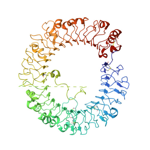
3W3K

Crystal structure of human TLR8 in complex with CL075


Experimental Data Snapshot

  • Method: X-RAY DIFFRACTION
  • Resolution: 2.30 Å
  • R-Value Free: 0.244 
  • R-Value Work: 0.205 
  • R-Value Observed: 0.207 

wwPDB Validation   3D Report Full Report


Ligand Structure Quality Assessment 


This is version 2.1 of the entry. See complete history


Literature

Structural reorganization of the Toll-like receptor 8 dimer induced by agonistic ligands

Tanji, H.Ohto, U.Shibata, T.Miyake, K.Shimizu, T.

(2013) Science 339: 1426-1429

  • DOI: https://doi.org/10.1126/science.1229159
  • Primary Citation of Related Structures:  
    3W3G, 3W3J, 3W3K, 3W3L, 3W3M, 3W3N

  • PubMed Abstract: 

    Toll-like receptor 7 (TLR7) and TLR8 recognize single-stranded RNA and initiate innate immune responses. Several synthetic agonists of TLR7-TLR8 display novel therapeutic potential; however, the molecular basis for ligand recognition and activation of signaling by TLR7 or TLR8 is largely unknown. In this study, the crystal structures of unliganded and ligand-induced activated human TLR8 dimers were elucidated. Ligand recognition was mediated by a dimerization interface formed by two protomers. Upon ligand stimulation, the TLR8 dimer was reorganized such that the two C termini were brought into proximity. The loop between leucine-rich repeat 14 (LRR14) and LRR15 was cleaved; however, the N- and C-terminal halves remained associated and contributed to ligand recognition and dimerization. Thus, ligand binding induces reorganization of the TLR8 dimer, which enables downstream signaling processes.


  • Organizational Affiliation

    Graduate School of Pharmaceutical Sciences, The University of Tokyo, Hongo, Bunkyo-ku, Tokyo 113-0033, Japan.


Macromolecules
Find similar proteins by:  (by identity cutoff)  |  3D Structure
Entity ID: 1
MoleculeChains Sequence LengthOrganismDetailsImage
Toll-like receptor 8
A, B
811Homo sapiensMutation(s): 0 
Gene Names: TLR8UNQ249/PRO286
UniProt & NIH Common Fund Data Resources
Find proteins for Q9NR97 (Homo sapiens)
Explore Q9NR97 
Go to UniProtKB:  Q9NR97
PHAROS:  Q9NR97
GTEx:  ENSG00000101916 
Entity Groups  
Sequence Clusters30% Identity50% Identity70% Identity90% Identity95% Identity100% Identity
UniProt GroupQ9NR97
Glycosylation
Glycosylation Sites: 6Go to GlyGen: Q9NR97-1
Sequence Annotations
Expand
  • Reference Sequence
Oligosaccharides

Help

Entity ID: 2
MoleculeChains Length2D Diagram Glycosylation3D Interactions
alpha-D-mannopyranose-(1-3)-[alpha-D-mannopyranose-(1-6)]beta-D-mannopyranose-(1-4)-2-acetamido-2-deoxy-beta-D-glucopyranose-(1-4)-2-acetamido-2-deoxy-beta-D-glucopyranose
C, E, F, H
5N-Glycosylation
Glycosylation Resources
GlyTouCan:  G22768VO
GlyCosmos:  G22768VO
GlyGen:  G22768VO
Entity ID: 3
MoleculeChains Length2D Diagram Glycosylation3D Interactions
beta-D-mannopyranose-(1-4)-2-acetamido-2-deoxy-beta-D-glucopyranose-(1-4)-2-acetamido-2-deoxy-beta-D-glucopyranose
D
3N-Glycosylation
Glycosylation Resources
GlyTouCan:  G15407YE
GlyCosmos:  G15407YE
GlyGen:  G15407YE
Entity ID: 4
MoleculeChains Length2D Diagram Glycosylation3D Interactions
2-acetamido-2-deoxy-beta-D-glucopyranose-(1-4)-2-acetamido-2-deoxy-beta-D-glucopyranose
G
2N-Glycosylation
Glycosylation Resources
GlyTouCan:  G42666HT
GlyCosmos:  G42666HT
GlyGen:  G42666HT
Small Molecules
Ligands 2 Unique
IDChains Name / Formula / InChI Key2D Diagram3D Interactions
L07
Query on L07

Download Ideal Coordinates CCD File 
I [auth A],
M [auth B]
2-propyl[1,3]thiazolo[4,5-c]quinolin-4-amine
C13 H13 N3 S
NFYMGJSUKCDVJR-UHFFFAOYSA-N
NAG
Query on NAG

Download Ideal Coordinates CCD File 
J [auth A]
K [auth A]
L [auth A]
N [auth B]
O [auth B]
J [auth A],
K [auth A],
L [auth A],
N [auth B],
O [auth B],
P [auth B]
2-acetamido-2-deoxy-beta-D-glucopyranose
C8 H15 N O6
OVRNDRQMDRJTHS-FMDGEEDCSA-N
Binding Affinity Annotations 
IDSourceBinding Affinity
L07 BindingDB:  3W3K EC50: min: 800, max: 4900 (nM) from 3 assay(s)
Experimental Data & Validation

Experimental Data

  • Method: X-RAY DIFFRACTION
  • Resolution: 2.30 Å
  • R-Value Free: 0.244 
  • R-Value Work: 0.205 
  • R-Value Observed: 0.207 
  • Space Group: P 1 21 1
Unit Cell:
Length ( Å )Angle ( ˚ )
a = 72.41α = 90
b = 154.02β = 103.03
c = 86.86γ = 90
Software Package:
Software NamePurpose
PHENIXrefinement
PDB_EXTRACTdata extraction
MOSFLMdata reduction
SCALAdata scaling
MOLREPphasing

Structure Validation

View Full Validation Report


Ligand Structure Quality Assessment 


Entry History 

Deposition Data

Revision History  (Full details and data files)

  • Version 1.0: 2013-04-03
    Type: Initial release
  • Version 2.0: 2020-07-29
    Type: Remediation
    Reason: Carbohydrate remediation
    Changes: Advisory, Atomic model, Data collection, Database references, Derived calculations, Structure summary
  • Version 2.1: 2024-10-16
    Changes: Data collection, Database references, Structure summary