3W9J

Structural basis for the inhibition of bacterial multidrug exporters


Experimental Data Snapshot

  • Method: X-RAY DIFFRACTION
  • Resolution: 3.15 Å
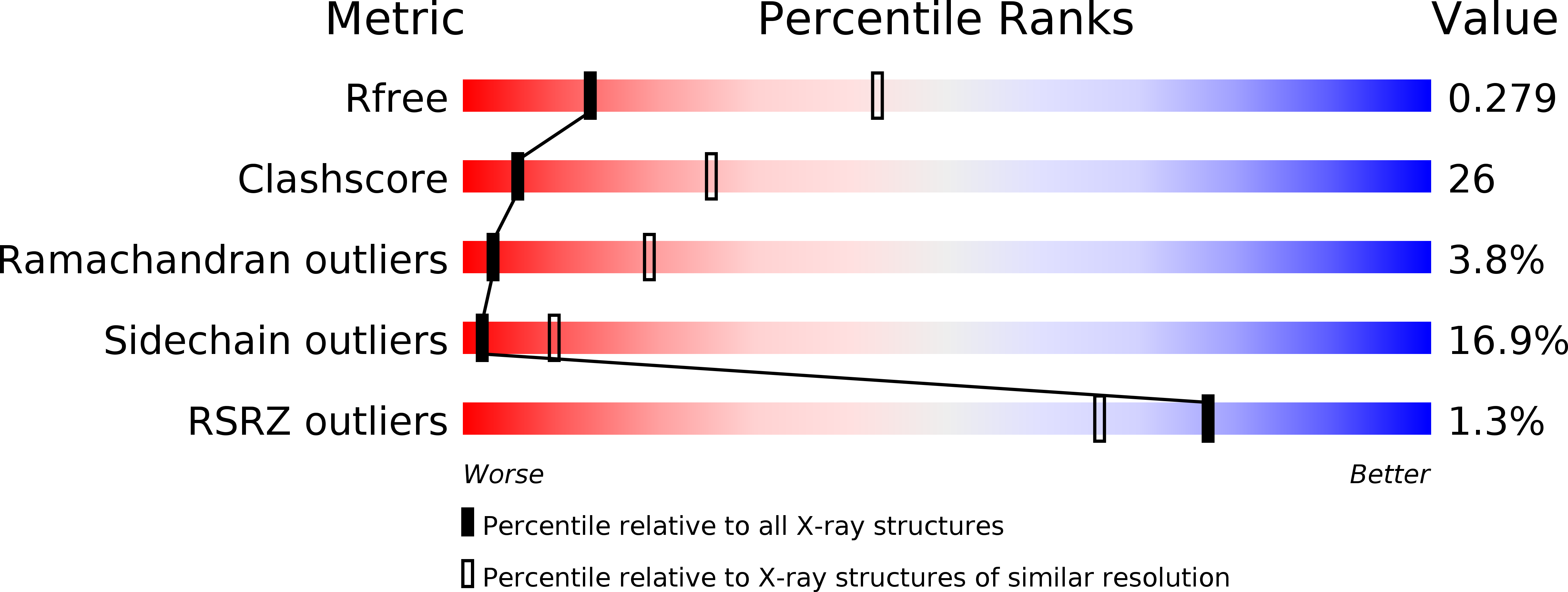
  • R-Value Free: 0.279 
  • R-Value Work: 0.202 
  • R-Value Observed: 0.206 

Starting Model: experimental
View more details

wwPDB Validation   3D Report Full Report


Ligand Structure Quality Assessment 


This is version 1.3 of the entry. See complete history


Literature

Structural basis for the inhibition of bacterial multidrug exporters

Nakashima, R.Sakurai, K.Yamasaki, S.Hayashi, K.Nagata, C.Hoshino, K.Onodera, Y.Nishino, K.Yamaguchi, A.

(2013) Nature 500: 102-106

  • DOI: https://doi.org/10.1038/nature12300
  • Primary Citation of Related Structures:  
    3W9H, 3W9I, 3W9J

  • PubMed Abstract: 

    The multidrug efflux transporter AcrB and its homologues are important in the multidrug resistance of Gram-negative pathogens. However, despite efforts to develop efflux inhibitors, clinically useful inhibitors are not available at present. Pyridopyrimidine derivatives are AcrB- and MexB-specific inhibitors that do not inhibit MexY; MexB and MexY are principal multidrug exporters in Pseudomonas aeruginosa. We have previously determined the crystal structure of AcrB in the absence and presence of antibiotics. Drugs were shown to be exported by a functionally rotating mechanism through tandem proximal and distal multisite drug-binding pockets. Here we describe the first inhibitor-bound structures of AcrB and MexB, in which these proteins are bound by a pyridopyrimidine derivative. The pyridopyrimidine derivative binds tightly to a narrow pit composed of a phenylalanine cluster located in the distal pocket and sterically hinders the functional rotation. This pit is a hydrophobic trap that branches off from the substrate-translocation channel. Phe 178 is located at the edge of this trap in AcrB and MexB and contributes to the tight binding of the inhibitor molecule through a π-π interaction with the pyridopyrimidine ring. The voluminous side chain of Trp 177 located at the corresponding position in MexY prevents inhibitor binding. The structure of the hydrophobic trap described in this study will contribute to the development of universal inhibitors of MexB and MexY in P. aeruginosa.


  • Organizational Affiliation

    Department of Cell Membrane Biology, Institute of Scientific and Industrial Research, Osaka University, Ibaraki, Osaka 567-0047, Japan.


Macromolecules
Find similar proteins by:  (by identity cutoff)  |  3D Structure
Entity ID: 1
MoleculeChains Sequence LengthOrganismDetailsImage
Multidrug resistance protein MexB
A, B, C, D, E
A, B, C, D, E, F
1,052Pseudomonas aeruginosa PAO1Mutation(s): 0 
Gene Names: mexBPA0426
Membrane Entity: Yes 
UniProt
Find proteins for P52002 (Pseudomonas aeruginosa (strain ATCC 15692 / DSM 22644 / CIP 104116 / JCM 14847 / LMG 12228 / 1C / PRS 101 / PAO1))
Explore P52002 
Go to UniProtKB:  P52002
Entity Groups  
Sequence Clusters30% Identity50% Identity70% Identity90% Identity95% Identity100% Identity
UniProt GroupP52002
Sequence Annotations
Expand
  • Reference Sequence
Small Molecules
Ligands 2 Unique
IDChains Name / Formula / InChI Key2D Diagram3D Interactions
P9D
Query on P9D

Download Ideal Coordinates CCD File 
J [auth B],
R [auth E]
[{2-[({[(3R)-1-{8-[(4-tert-butyl-1,3-thiazol-2-yl)carbamoyl]-4-oxo-3-[(E)-2-(1H-tetrazol-5-yl)ethenyl]-4H-pyrido[1,2-a]pyrimidin-2-yl}piperidin-3-yl]oxy}carbonyl)amino]ethyl}(dimethyl)ammonio]acetate
C31 H39 N11 O6 S
RVJNIKFVEAWLQC-HXUWFJFHSA-N
LMT
Query on LMT

Download Ideal Coordinates CCD File 
G [auth A]
H [auth A]
I [auth A]
K [auth B]
L [auth B]
G [auth A],
H [auth A],
I [auth A],
K [auth B],
L [auth B],
M [auth B],
N [auth C],
O [auth D],
P [auth D],
Q [auth D],
S [auth E],
T [auth E],
U [auth E],
V [auth F],
W [auth F]
DODECYL-BETA-D-MALTOSIDE
C24 H46 O11
NLEBIOOXCVAHBD-QKMCSOCLSA-N
Experimental Data & Validation

Experimental Data

  • Method: X-RAY DIFFRACTION
  • Resolution: 3.15 Å
  • R-Value Free: 0.279 
  • R-Value Work: 0.202 
  • R-Value Observed: 0.206 
  • Space Group: P 1
Unit Cell:
Length ( Å )Angle ( ˚ )
a = 126.204α = 85.75
b = 137.034β = 68.93
c = 152.275γ = 87.39
Software Package:
Software NamePurpose
DENZOdata reduction
SCALEPACKdata scaling
MOLREPphasing
REFMACrefinement
PDB_EXTRACTdata extraction

Structure Validation

View Full Validation Report


Ligand Structure Quality Assessment 


Entry History 

Deposition Data

Revision History  (Full details and data files)

  • Version 1.0: 2013-07-03
    Type: Initial release
  • Version 1.1: 2013-08-07
    Changes: Database references
  • Version 1.2: 2017-11-22
    Changes: Refinement description
  • Version 1.3: 2023-11-08
    Changes: Data collection, Database references, Derived calculations, Refinement description