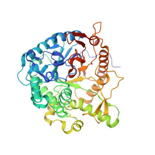
3WH7

Crystal structure of GH1 beta-glucosidase Td2F2 L-fucose complex


Experimental Data Snapshot

  • Method: X-RAY DIFFRACTION
  • Resolution: 1.10 Å
  • R-Value Free: 0.134 
  • R-Value Work: 0.113 
  • R-Value Observed: 0.114 

Starting Model: experimental
View more details

wwPDB Validation   3D Report Full Report


Ligand Structure Quality Assessment 


This is version 1.4 of the entry. See complete history


Literature

Crystal structure and identification of a key amino acid for glucose tolerance, substrate specificity, and transglycosylation activity of metagenomic beta-glucosidase Td2F2.

Matsuzawa, T.Jo, T.Uchiyama, T.Manninen, J.A.Arakawa, T.Miyazaki, K.Fushinobu, S.Yaoi, K.

(2016) FEBS J 283: 2340-2353

  • DOI: https://doi.org/10.1111/febs.13743
  • Primary Citation of Related Structures:  
    3WH5, 3WH6, 3WH7, 3WH8, 5AYB, 5AYI

  • PubMed Abstract: 

    β-Glucosidase Td2F2 isolated from a compost metagenome has high glucose tolerance and transglycosylation activity. In this study, we determined the high-resolution crystal structure of Td2F2. It has a unique structure at the -1 subsite that is important for substrate specificity but not for glucose tolerance. To elucidate the mechanism(s) of glucose tolerance, we isolated a glucose-sensitive Td2F2 mutant using random mutagenesis. In this mutant, Asn223 residue located between subsites +1 and +2 was mutated. The Asn223 mutation resulted in reduced glucose tolerance and transglycosylation activity, and drastically changed substrate specificity. These results indicate that the structure between subsites +1 and +2 is critical for the glucose tolerance and substrate specificity of Td2F2. Our findings shed light on the glucose tolerance and transglycosylation activity mechanisms of glycoside hydrolase family 1 β-glucosidases. The atomic coordinates and structure factors (codes 3WH5, 3WH6, 3WH8, 3WH7, 5AYB, and 5AYI) have been deposited in the Protein Data Bank (http://wwpdb.org/).


  • Organizational Affiliation

    Bioproduction Research Institute, National Institute of Advanced Industrial Science and Technology (AIST), Tsukuba, Ibaraki, Japan.


Macromolecules
Find similar proteins by:  (by identity cutoff)  |  3D Structure
Entity ID: 1
MoleculeChains Sequence LengthOrganismDetailsImage
beta-glucosidase457metagenomesMutation(s): 0 
EC: 3.2.1.21
Entity Groups  
Sequence Clusters30% Identity50% Identity70% Identity90% Identity95% Identity100% Identity
Sequence Annotations
Expand
  • Reference Sequence
Experimental Data & Validation

Experimental Data

  • Method: X-RAY DIFFRACTION
  • Resolution: 1.10 Å
  • R-Value Free: 0.134 
  • R-Value Work: 0.113 
  • R-Value Observed: 0.114 
  • Space Group: P 21 21 21
Unit Cell:
Length ( Å )Angle ( ˚ )
a = 69.05α = 90
b = 69.693β = 90
c = 96.209γ = 90
Software Package:
Software NamePurpose
HKL-2000data collection
MOLREPphasing
REFMACrefinement
HKL-2000data reduction
HKL-2000data scaling

Structure Validation

View Full Validation Report


Ligand Structure Quality Assessment 


Entry History 

Deposition Data

Revision History  (Full details and data files)

  • Version 1.0: 2014-09-03
    Type: Initial release
  • Version 1.1: 2016-05-04
    Changes: Database references
  • Version 1.2: 2020-07-29
    Type: Remediation
    Reason: Carbohydrate remediation
    Changes: Data collection, Derived calculations, Structure summary
  • Version 1.3: 2022-08-24
    Changes: Database references, Structure summary
  • Version 1.4: 2023-11-08
    Changes: Data collection, Refinement description