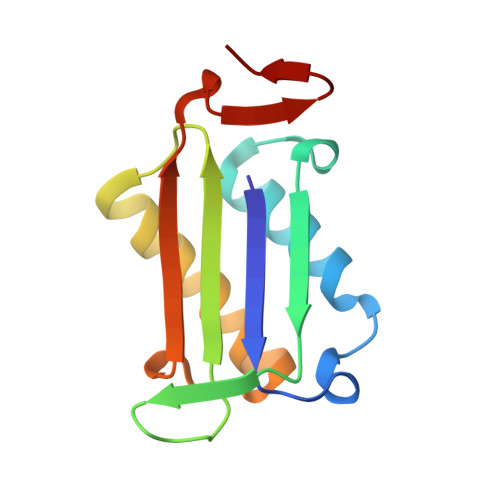
3WNT

Multiple binding modes of benzyl isothiocyanate inhibitor complexed with Macrophage Migration Inhibitory Factor


Experimental Data Snapshot

  • Method: X-RAY DIFFRACTION
  • Resolution: 2.07 Å
  • R-Value Free: 0.263 
  • R-Value Work: 0.222 
  • R-Value Observed: 0.224 

wwPDB Validation   3D Report Full Report


Ligand Structure Quality Assessment 


This is version 1.1 of the entry. See complete history


Literature

Multiple binding modes of isothiocyanates that inhibit macrophage migration inhibitory factor

Spencer, E.S.Dale, E.J.Gommans, A.L.Rutledge, M.T.Vo, C.T.Nakatani, Y.Gamble, A.B.Smith, R.A.Wilbanks, S.M.Hampton, M.B.Tyndall, J.D.

(2015) Eur J Med Chem 93: 501-510

  • DOI: https://doi.org/10.1016/j.ejmech.2015.02.012
  • Primary Citation of Related Structures:  
    3WNR, 3WNS, 3WNT, 4OSF

  • PubMed Abstract: 

    Macrophage migration inhibitory factor (MIF) is a pleiotropic cytokine that has roles in the innate immune response, and also contributes to inflammatory disease. While the biological properties of MIF are closely linked to protein-protein interactions, MIF also has tautomerase activity. Inhibition of this activity interferes with the interaction of MIF with protein partners e.g. the CD74 receptor, and tautomerase inhibitors show promise in disease models including multiple sclerosis and colitis. Isothiocyanates inhibit MIF tautomerase activity via covalent modification of the N-terminal proline. We systematically explored variants of benzyl and phenethyl isothiocyanates, to define determinants of inhibition. In particular, substitution with hydroxyl, chloro, fluoro and trifluoro moieties at the para and meta positions were evaluated. In assays on treated cells and recombinant protein, the IC50 varied from 250 nM to >100 μM. X-ray crystal structures of selected complexes revealed that two binding modes are accessed by some compounds, perhaps owing to strain in short linkers between the isothiocyanate and aromatic ring. The variety of binding modes confirms the existence of two subsites for inhibitors and establishes a platform for the development of potent inhibitors of MIF that only need to target one of these subsites.


  • Organizational Affiliation

    Centre for Free Radical Research, Department of Pathology, University of Otago, PO Box 4345, Christchurch 8140, New Zealand.


Macromolecules
Find similar proteins by:  (by identity cutoff)  |  3D Structure
Entity ID: 1
MoleculeChains Sequence LengthOrganismDetailsImage
Macrophage migration inhibitory factor
A, B, C
114Homo sapiensMutation(s): 0 
Gene Names: GLIFMIFMMIF
EC: 5.3.2.1 (PDB Primary Data), 5.3.3.12 (PDB Primary Data)
UniProt & NIH Common Fund Data Resources
Find proteins for P14174 (Homo sapiens)
Explore P14174 
Go to UniProtKB:  P14174
PHAROS:  P14174
GTEx:  ENSG00000240972 
Entity Groups  
Sequence Clusters30% Identity50% Identity70% Identity90% Identity95% Identity100% Identity
UniProt GroupP14174
Sequence Annotations
Expand
  • Reference Sequence
Small Molecules
Ligands 3 Unique
IDChains Name / Formula / InChI Key2D Diagram3D Interactions
9BE
Query on 9BE

Download Ideal Coordinates CCD File 
D [auth A],
G [auth B],
I [auth C]
N-benzylthioformamide
C8 H9 N S
QAADZYUXQLUXFX-UHFFFAOYSA-N
SO4
Query on SO4

Download Ideal Coordinates CCD File 
E [auth A],
F [auth A],
H [auth B],
J [auth C],
K [auth C]
SULFATE ION
O4 S
QAOWNCQODCNURD-UHFFFAOYSA-L
IPA
Query on IPA

Download Ideal Coordinates CCD File 
L [auth C]ISOPROPYL ALCOHOL
C3 H8 O
KFZMGEQAYNKOFK-UHFFFAOYSA-N
Experimental Data & Validation

Experimental Data

  • Method: X-RAY DIFFRACTION
  • Resolution: 2.07 Å
  • R-Value Free: 0.263 
  • R-Value Work: 0.222 
  • R-Value Observed: 0.224 
  • Space Group: P 21 21 21
Unit Cell:
Length ( Å )Angle ( ˚ )
a = 68.17α = 90
b = 68.56β = 90
c = 88.67γ = 90
Software Package:
Software NamePurpose
SCALAdata scaling
PHASERphasing
PHENIXrefinement
PDB_EXTRACTdata extraction
Blu-Icedata collection
MOSFLMdata reduction

Structure Validation

View Full Validation Report


Ligand Structure Quality Assessment 


Entry History 

Deposition Data

Revision History  (Full details and data files)

  • Version 1.0: 2014-03-05
    Type: Initial release
  • Version 1.1: 2015-04-01
    Changes: Database references