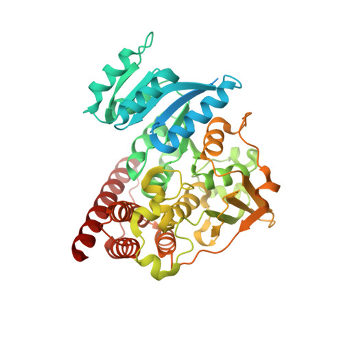
3WQQ

Crystal structure of PfDXR complexed with inhibitor-3


Experimental Data Snapshot

  • Method: X-RAY DIFFRACTION
  • Resolution: 2.25 Å
  • R-Value Free: 0.240 
  • R-Value Work: 0.185 
  • R-Value Observed: 0.188 

wwPDB Validation   3D Report Full Report


Ligand Structure Quality Assessment 


This is version 1.1 of the entry. See complete history


Literature

Binding Modes of Reverse Fosmidomycin Analogs toward the Antimalarial Target IspC.

Konzuch, S.Umeda, T.Held, J.Hahn, S.Brucher, K.Lienau, C.Behrendt, C.T.Grawert, T.Bacher, A.Illarionov, B.Fischer, M.Mordmuller, B.Tanaka, N.Kurz, T.

(2014) J Med Chem 57: 8827-8838

  • DOI: https://doi.org/10.1021/jm500850y
  • Primary Citation of Related Structures:  
    3WQQ, 3WQR, 3WQS

  • PubMed Abstract: 

    1-Deoxy-d-xylulose 5-phosphate reductoisomerase of Plasmodium falciparum (PfIspC, PfDxr), believed to be the rate-limiting enzyme of the nonmevalonate pathway of isoprenoid biosynthesis (MEP pathway), is a clinically validated antimalarial target. The enzyme is efficiently inhibited by the natural product fosmidomycin. To gain new insights into the structure activity relationships of reverse fosmidomycin analogs, several reverse analogs of fosmidomycin were synthesized and biologically evaluated. The 4-methoxyphenyl substituted derivative 2c showed potent inhibition of PfIspC as well as of P. falciparum growth and was more than one order of magnitude more active than fosmidomycin. The binding modes of three new derivatives in complex with PfIspC, reduced nicotinamide adenine dinucleotide phosphate, and Mg(2+) were determined by X-ray structure analysis. Notably, PfIspC selectively binds the S-enantiomers of the study compounds.


  • Organizational Affiliation

    Institut für Pharmazeutische und Medizinische Chemie, Heinrich Heine Universität , Universitätsstr. 1, 40225 Düsseldorf, Germany.


Macromolecules
Find similar proteins by:  (by identity cutoff)  |  3D Structure
Entity ID: 1
MoleculeChains Sequence LengthOrganismDetailsImage
1-deoxy-D-xylulose 5-phosphate reductoisomerase, apicoplast
A, B
488Plasmodium falciparum HB3Mutation(s): 0 
Gene Names: DXR
EC: 1.1.1.267
UniProt
Find proteins for O96693 (Plasmodium falciparum (isolate HB3))
Explore O96693 
Go to UniProtKB:  O96693
Entity Groups  
Sequence Clusters30% Identity50% Identity70% Identity90% Identity95% Identity100% Identity
UniProt GroupO96693
Sequence Annotations
Expand
  • Reference Sequence
Small Molecules
Ligands 4 Unique
IDChains Name / Formula / InChI Key2D Diagram3D Interactions
NDP
Query on NDP

Download Ideal Coordinates CCD File 
C [auth A],
F [auth B]
NADPH DIHYDRO-NICOTINAMIDE-ADENINE-DINUCLEOTIDE PHOSPHATE
C21 H30 N7 O17 P3
ACFIXJIJDZMPPO-NNYOXOHSSA-N
IB3
Query on IB3

Download Ideal Coordinates CCD File 
E [auth A],
J [auth B]
[(1S)-4-[hydroxy(methyl)amino]-1-(4-methylphenyl)-4-oxobutyl]phosphonic acid
C12 H18 N O5 P
YBVPWURHYFIQDS-NSHDSACASA-N
CA
Query on CA

Download Ideal Coordinates CCD File 
H [auth B],
I [auth B]
CALCIUM ION
Ca
BHPQYMZQTOCNFJ-UHFFFAOYSA-N
MG
Query on MG

Download Ideal Coordinates CCD File 
D [auth A],
G [auth B]
MAGNESIUM ION
Mg
JLVVSXFLKOJNIY-UHFFFAOYSA-N
Experimental Data & Validation

Experimental Data

  • Method: X-RAY DIFFRACTION
  • Resolution: 2.25 Å
  • R-Value Free: 0.240 
  • R-Value Work: 0.185 
  • R-Value Observed: 0.188 
  • Space Group: P 1 21 1
Unit Cell:
Length ( Å )Angle ( ˚ )
a = 51.042α = 90
b = 76.26β = 90.82
c = 104.231γ = 90
Software Package:
Software NamePurpose
REFMACrefinement
HKL-2000data reduction
HKL-2000data scaling

Structure Validation

View Full Validation Report


Ligand Structure Quality Assessment 


Entry History 

Deposition Data

Revision History  (Full details and data files)

  • Version 1.0: 2014-11-26
    Type: Initial release
  • Version 1.1: 2024-03-20
    Changes: Data collection, Database references, Derived calculations