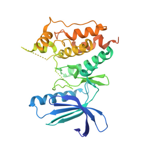
3WYX

CRYSTAL STRUCTURE OF HUMAN MPS1 CATALYTIC DOMAIN IN COMPLEX WITH 6-((3-(cyanomethoxy)-4-(1-methyl-1H-pyrazol-4-yl)phenyl)amino)-2-(cyclohexylamino)nicotinonitrile


Experimental Data Snapshot

  • Method: X-RAY DIFFRACTION
  • Resolution: 2.90 Å
  • R-Value Free: 0.273 
  • R-Value Work: 0.232 
  • R-Value Observed: 0.234 

wwPDB Validation   3D Report Full Report


Ligand Structure Quality Assessment 


This is version 1.2 of the entry. See complete history


Literature

A unique hinge binder of extremely selective aminopyridine-based Mps1 (TTK) kinase inhibitors with cellular activity.

Kusakabe, K.Ide, N.Daigo, Y.Itoh, T.Yamamoto, T.Kojima, E.Mitsuoka, Y.Tadano, G.Tagashira, S.Higashino, K.Okano, Y.Sato, Y.Inoue, M.Iguchi, M.Kanazawa, T.Ishioka, Y.Dohi, K.Kido, Y.Sakamoto, S.Ando, S.Maeda, M.Higaki, M.Yoshizawa, H.Murai, H.Nakamura, Y.

(2015) Bioorg Med Chem 23: 2247-2260

  • DOI: https://doi.org/10.1016/j.bmc.2015.02.042
  • Primary Citation of Related Structures:  
    3WYX, 3WYY

  • PubMed Abstract: 

    Mps1, also known as TTK, is a dual-specificity kinase that regulates the spindle assembly check point. Increased expression levels of Mps1 are observed in cancer cells, and the expression levels correlate well with tumor grade. Such evidence points to selective inhibition of Mps1 as an attractive strategy for cancer therapeutics. Starting from an aminopyridine-based lead 3a that binds to a flipped-peptide conformation at the hinge region in Mps1, elaboration of the aminopyridine scaffold at the 2- and 6-positions led to the discovery of 19c that exhibited no significant inhibition for 287 kinases as well as improved cellular Mps1 and antiproliferative activities in A549 lung carcinoma cells (cellular Mps1 IC₅₀=5.3 nM, A549 IC₅₀=26 nM). A clear correlation between cellular Mps1 and antiproliferative IC₅₀ values indicated that the antiproliferative activity observed in A549 cells would be responsible for the cellular inhibition of Mps1. The X-ray structure of 19c in complex with Mps1 revealed that this compound retains the ability to bind to the peptide flip conformation. Finally, comparative analysis of the X-ray structures of 19c, a deamino analogue 33, and a known Mps1 inhibitor bound to Mps1 provided insights into the unique binding mode at the hinge region.


  • Organizational Affiliation

    Medicinal Research Laboratories, Shionogi Pharmaceutical Research Center, 1-1 Futaba-cho 3-chome, Toyonaka, Osaka 561-0825, Japan. Electronic address: [email protected].


Macromolecules
Find similar proteins by:  (by identity cutoff)  |  3D Structure
Entity ID: 1
MoleculeChains Sequence LengthOrganismDetailsImage
Dual specificity protein kinase TTK321Homo sapiensMutation(s): 0 
Gene Names: TTKMPS1MPS1L1
EC: 2.7.12.1
UniProt & NIH Common Fund Data Resources
Find proteins for P33981 (Homo sapiens)
Explore P33981 
Go to UniProtKB:  P33981
PHAROS:  P33981
GTEx:  ENSG00000112742 
Entity Groups  
Sequence Clusters30% Identity50% Identity70% Identity90% Identity95% Identity100% Identity
UniProt GroupP33981
Sequence Annotations
Expand
  • Reference Sequence
Small Molecules
Ligands 2 Unique
IDChains Name / Formula / InChI Key2D Diagram3D Interactions
O38
Query on O38

Download Ideal Coordinates CCD File 
C [auth A]6-{[3-(cyanomethoxy)-4-(1-methyl-1H-pyrazol-4-yl)phenyl]amino}-2-(cyclohexylamino)pyridine-3-carbonitrile
C24 H25 N7 O
OVHGMKGIRAADEL-UHFFFAOYSA-N
IOD
Query on IOD

Download Ideal Coordinates CCD File 
B [auth A]IODIDE ION
I
XMBWDFGMSWQBCA-UHFFFAOYSA-M
Experimental Data & Validation

Experimental Data

  • Method: X-RAY DIFFRACTION
  • Resolution: 2.90 Å
  • R-Value Free: 0.273 
  • R-Value Work: 0.232 
  • R-Value Observed: 0.234 
  • Space Group: I 2 2 2
Unit Cell:
Length ( Å )Angle ( ˚ )
a = 70.25α = 90
b = 105.57β = 90
c = 113.38γ = 90
Software Package:
Software NamePurpose
MOSFLMdata reduction
SCALAdata scaling
MOLREPphasing
REFMACrefinement
PDB_EXTRACTdata extraction
CrystalCleardata collection

Structure Validation

View Full Validation Report


Ligand Structure Quality Assessment 


Entry History 

Revision History  (Full details and data files)

  • Version 1.0: 2015-04-08
    Type: Initial release
  • Version 1.1: 2022-08-24
    Changes: Database references, Derived calculations
  • Version 1.2: 2024-05-29
    Changes: Data collection