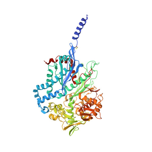
4A5A

Crystal structure of the C258S/C268S variant of Toxoplasma gondii nucleoside triphosphate diphosphohydrolase 3 (NTPDase3) in complex with magnesium and AMPPNP


Experimental Data Snapshot

  • Method: X-RAY DIFFRACTION
  • Resolution: 2.85 Å
  • R-Value Free: 0.284 
  • R-Value Work: 0.234 
  • R-Value Observed: 0.236 

Starting Model: experimental
View more details

wwPDB Validation   3D Report Full Report


Ligand Structure Quality Assessment 


This is version 1.5 of the entry. See complete history


Literature

Structural Insight Into the Activation Mechanism of Toxoplasma Gondii Nucleoside Triphosphate Diphosphohydrolases by Disulfide Reduction.

Krug, U.Zebisch, M.Krauss, M.Straeter, N.

(2012) J Biol Chem 287: 3051

  • DOI: https://doi.org/10.1074/jbc.M111.294348
  • Primary Citation of Related Structures:  
    4A57, 4A59, 4A5A, 4A5B

  • PubMed Abstract: 

    The intracellular parasite Toxoplasma gondii produces two nucleoside triphosphate diphosphohydrolases (NTPDase1 and -3). These tetrameric, cysteine-rich enzymes require activation by reductive cleavage of a hitherto unknown disulfide bond. Despite a 97% sequence identity, both isozymes differ largely in their ability to hydrolyze ATP and ADP. Here, we present crystal structures of inactive NTPDase3 as an apo form and in complex with the product AMP to resolutions of 2.0 and 2.2 Å, respectively. We find that the enzyme is present in an open conformation that precludes productive substrate binding and catalysis. The cysteine bridge 258-268 is identified to be responsible for locking of activity. Crystal structures of constitutively active variants of NTPDase1 and -3 generated by mutation of Cys(258)-Cys(268) show that opening of the regulatory cysteine bridge induces a pronounced contraction of the whole tetramer. This is accompanied by a 12° domain closure motion resulting in the correct arrangement of all active site residues. A complex structure of activated NTPDase3 with a non-hydrolyzable ATP analog and the cofactor Mg(2+) to a resolution of 2.85 Å indicates that catalytic differences between the NTPDases are primarily dictated by differences in positioning of the adenine base caused by substitution of Arg(492) and Glu(493) in NTPDase1 by glycines in NTPDase3.


  • Organizational Affiliation

    Institute of Bioanalytical Chemistry, Center for Biotechnology and Biomedicine, University of Leipzig, D-04103 Leipzig, Germany.


Macromolecules
Find similar proteins by:  (by identity cutoff)  |  3D Structure
Entity ID: 1
MoleculeChains Sequence LengthOrganismDetailsImage
NUCLEOSIDE-TRIPHOSPHATASE 1
A, B, C, D
611Toxoplasma gondiiMutation(s): 2 
EC: 3.6.1.5 (PDB Primary Data), 3.6.1.15 (UniProt)
UniProt
Find proteins for Q27893 (Toxoplasma gondii)
Explore Q27893 
Go to UniProtKB:  Q27893
Entity Groups  
Sequence Clusters30% Identity50% Identity70% Identity90% Identity95% Identity100% Identity
UniProt GroupQ27893
Sequence Annotations
Expand
  • Reference Sequence
Small Molecules
Ligands 2 Unique
IDChains Name / Formula / InChI Key2D Diagram3D Interactions
ANP
Query on ANP

Download Ideal Coordinates CCD File 
E [auth A],
G [auth B],
I [auth C],
K [auth D]
PHOSPHOAMINOPHOSPHONIC ACID-ADENYLATE ESTER
C10 H17 N6 O12 P3
PVKSNHVPLWYQGJ-KQYNXXCUSA-N
MG
Query on MG

Download Ideal Coordinates CCD File 
F [auth A],
H [auth B],
J [auth C],
L [auth D]
MAGNESIUM ION
Mg
JLVVSXFLKOJNIY-UHFFFAOYSA-N
Experimental Data & Validation

Experimental Data

  • Method: X-RAY DIFFRACTION
  • Resolution: 2.85 Å
  • R-Value Free: 0.284 
  • R-Value Work: 0.234 
  • R-Value Observed: 0.236 
  • Space Group: C 2 2 21
Unit Cell:
Length ( Å )Angle ( ˚ )
a = 71.953α = 90
b = 150.668β = 90
c = 486.988γ = 90
Software Package:
Software NamePurpose
REFMACrefinement
XDSdata reduction
SCALAdata scaling
PHASERphasing

Structure Validation

View Full Validation Report


Ligand Structure Quality Assessment 


Entry History 

Deposition Data

Revision History  (Full details and data files)

  • Version 1.0: 2011-11-30
    Type: Initial release
  • Version 1.1: 2012-01-11
    Changes: Other
  • Version 1.2: 2012-02-15
    Changes: Other
  • Version 1.3: 2013-12-25
    Changes: Structure summary
  • Version 1.4: 2023-12-20
    Changes: Data collection, Database references, Derived calculations, Other, Refinement description
  • Version 1.5: 2024-11-06
    Changes: Structure summary