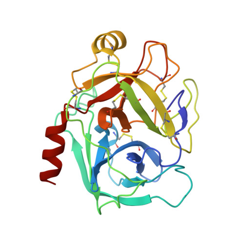
4ABG

Fragments bound to bovine trypsin for the SAMPL challenge


Experimental Data Snapshot

  • Method: X-RAY DIFFRACTION
  • Resolution: 1.52 Å
  • R-Value Free: 0.204 
  • R-Value Work: 0.165 
  • R-Value Observed: 0.167 

Starting Model: experimental
View more details

wwPDB Validation   3D Report Full Report


Ligand Structure Quality Assessment 


This is version 1.3 of the entry. See complete history


Literature

The Dingo Dataset: A Comprehensive Set of Data for the Sampl Challenge.

Newman, J.Dolezal, O.Fazio, V.Caradoc-Davies, T.Peat, T.S.

(2012) J Comput Aided Mol Des 26: 497

  • DOI: https://doi.org/10.1007/s10822-011-9521-2
  • Primary Citation of Related Structures:  
    4AB8, 4AB9, 4ABA, 4ABB, 4ABD, 4ABE, 4ABF, 4ABG, 4ABH

  • PubMed Abstract: 

    Part of the latest SAMPL challenge was to predict how a small fragment library of 500 commercially available compounds would bind to a protein target. In order to assess the modellers' work, a reasonably comprehensive set of data was collected using a number of techniques. These included surface plasmon resonance, isothermal titration calorimetry, protein crystallization and protein crystallography. Using these techniques we could determine the kinetics of fragment binding, the energy of binding, how this affects the ability of the target to crystallize, and when the fragment did bind, the pose or orientation of binding. Both the final data set and all of the raw images have been made available to the community for scrutiny and further work. This overview sets out to give the parameters of the experiments done and what might be done differently for future studies.


  • Organizational Affiliation

    CSIRO Division of Materials, Science and Engineering, 343 Royal Parade, Parkville, VIC 3052, Australia.


Macromolecules
Find similar proteins by:  (by identity cutoff)  |  3D Structure
Entity ID: 1
MoleculeChains Sequence LengthOrganismDetailsImage
CATIONIC TRYPSIN223Bos taurusMutation(s): 0 
EC: 3.4.21.4
UniProt
Find proteins for P00760 (Bos taurus)
Explore P00760 
Go to UniProtKB:  P00760
Entity Groups  
Sequence Clusters30% Identity50% Identity70% Identity90% Identity95% Identity100% Identity
UniProt GroupP00760
Sequence Annotations
Expand
  • Reference Sequence
Small Molecules
Ligands 5 Unique
IDChains Name / Formula / InChI Key2D Diagram3D Interactions
91B
Query on 91B

Download Ideal Coordinates CCD File 
K [auth A]1-[3-(4-METHYLPIPERAZIN-1-YL)PHENYL]METHANAMINE
C12 H19 N3
VKTLTRIKVYJVOX-UHFFFAOYSA-N
SO4
Query on SO4

Download Ideal Coordinates CCD File 
D [auth A],
E [auth A]
SULFATE ION
O4 S
QAOWNCQODCNURD-UHFFFAOYSA-L
EDO
Query on EDO

Download Ideal Coordinates CCD File 
F [auth A],
G [auth A],
H [auth A],
I [auth A],
J [auth A]
1,2-ETHANEDIOL
C2 H6 O2
LYCAIKOWRPUZTN-UHFFFAOYSA-N
CA
Query on CA

Download Ideal Coordinates CCD File 
B [auth A]CALCIUM ION
Ca
BHPQYMZQTOCNFJ-UHFFFAOYSA-N
CL
Query on CL

Download Ideal Coordinates CCD File 
C [auth A]CHLORIDE ION
Cl
VEXZGXHMUGYJMC-UHFFFAOYSA-M
Binding Affinity Annotations 
IDSourceBinding Affinity
91B PDBBind:  4ABG Kd: 2.71e+5 (nM) from 1 assay(s)
Experimental Data & Validation

Experimental Data

  • Method: X-RAY DIFFRACTION
  • Resolution: 1.52 Å
  • R-Value Free: 0.204 
  • R-Value Work: 0.165 
  • R-Value Observed: 0.167 
  • Space Group: P 31 2 1
Unit Cell:
Length ( Å )Angle ( ˚ )
a = 54.303α = 90
b = 54.303β = 90
c = 106.387γ = 120
Software Package:
Software NamePurpose
REFMACrefinement
XDSdata reduction
SCALAdata scaling
PHASERphasing

Structure Validation

View Full Validation Report


Ligand Structure Quality Assessment 


Entry History 

Deposition Data

Revision History  (Full details and data files)

  • Version 1.0: 2012-02-08
    Type: Initial release
  • Version 1.1: 2014-02-05
    Changes: Database references
  • Version 1.2: 2023-12-20
    Changes: Data collection, Database references, Derived calculations, Other, Refinement description
  • Version 1.3: 2024-11-13
    Changes: Structure summary