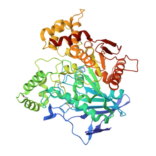
4BDS

Human butyrylcholinesterase in complex with tacrine


Experimental Data Snapshot

  • Method: X-RAY DIFFRACTION
  • Resolution: 2.10 Å
  • R-Value Free: 0.209 
  • R-Value Work: 0.175 
  • R-Value Observed: 0.176 

Starting Model: experimental
View more details

wwPDB Validation   3D Report Full Report


Ligand Structure Quality Assessment 


This is version 4.1 of the entry. See complete history


Literature

Crystal Structures of Human Cholinesterases in Complex with Huprine W and Tacrine: Elements of Specificity for Anti-Alzheimer'S Drugs Targeting Acetyl- and Butyrylcholinesterase.

Nachon, F.Carletti, E.Ronco, C.Trovaslet, M.Nicolet, Y.Jean, L.Renard, P.

(2013) Biochem J 453: 393

  • DOI: https://doi.org/10.1042/BJ20130013
  • Primary Citation of Related Structures:  
    4BDS, 4BDT

  • PubMed Abstract: 

    The multifunctional nature of Alzheimer's disease calls for MTDLs (multitarget-directed ligands) to act on different components of the pathology, like the cholinergic dysfunction and amyloid aggregation. Such MTDLs are usually on the basis of cholinesterase inhibitors (e.g. tacrine or huprine) coupled with another active molecule aimed at a different target. To aid in the design of these MTDLs, we report the crystal structures of hAChE (human acetylcholinesterase) in complex with FAS-2 (fasciculin 2) and a hydroxylated derivative of huprine (huprine W), and of hBChE (human butyrylcholinesterase) in complex with tacrine. Huprine W in hAChE and tacrine in hBChE reside in strikingly similar positions highlighting the conservation of key interactions, namely, π-π/cation-π interactions with Trp86 (Trp82), and hydrogen bonding with the main chain carbonyl of the catalytic histidine residue. Huprine W forms additional interactions with hAChE, which explains its superior affinity: the isoquinoline moiety is associated with a group of aromatic residues (Tyr337, Phe338 and Phe295 not present in hBChE) in addition to Trp86; the hydroxyl group is hydrogen bonded to both the catalytic serine residue and residues in the oxyanion hole; and the chlorine substituent is nested in a hydrophobic pocket interacting strongly with Trp439. There is no pocket in hBChE that is able to accommodate the chlorine substituent.


  • Organizational Affiliation

    Département de Toxicologie, Institut de Recherche Biomédicale des Armées, 24 Avenue des Maquis du Grésivaudan, BP87, 38702 La Tronche, France. [email protected]


Macromolecules
Find similar proteins by:  (by identity cutoff)  |  3D Structure
Entity ID: 1
MoleculeChains Sequence LengthOrganismDetailsImage
CHOLINESTERASE529Homo sapiensMutation(s): 4 
EC: 3.1.1.8
UniProt & NIH Common Fund Data Resources
Find proteins for P06276 (Homo sapiens)
Explore P06276 
Go to UniProtKB:  P06276
PHAROS:  P06276
GTEx:  ENSG00000114200 
Entity Groups  
Sequence Clusters30% Identity50% Identity70% Identity90% Identity95% Identity100% Identity
UniProt GroupP06276
Glycosylation
Glycosylation Sites: 6Go to GlyGen: P06276-1
Sequence Annotations
Expand
  • Reference Sequence
Oligosaccharides

Help

Entity ID: 2
MoleculeChains Length2D Diagram Glycosylation3D Interactions
2-acetamido-2-deoxy-beta-D-glucopyranose-(1-4)-[beta-L-fucopyranose-(1-6)]2-acetamido-2-deoxy-beta-D-glucopyranose
B, C
3N-Glycosylation
Glycosylation Resources
GlyTouCan:  G28454KX
GlyCosmos:  G28454KX
GlyGen:  G28454KX
Small Molecules
Ligands 8 Unique
IDChains Name / Formula / InChI Key2D Diagram3D Interactions
NAG
Query on NAG

Download Ideal Coordinates CCD File 
D [auth A],
E [auth A],
G [auth A],
H [auth A]
2-acetamido-2-deoxy-beta-D-glucopyranose
C8 H15 N O6
OVRNDRQMDRJTHS-FMDGEEDCSA-N
THA
Query on THA

Download Ideal Coordinates CCD File 
I [auth A]TACRINE
C13 H14 N2
YLJREFDVOIBQDA-UHFFFAOYSA-N
FUC
Query on FUC

Download Ideal Coordinates CCD File 
F [auth A]alpha-L-fucopyranose
C6 H12 O5
SHZGCJCMOBCMKK-SXUWKVJYSA-N
FPK
Query on FPK

Download Ideal Coordinates CCD File 
O [auth A]1-formyl-L-proline
C6 H9 N O3
DHDRGOURKDLAOT-YFKPBYRVSA-N
SO4
Query on SO4

Download Ideal Coordinates CCD File 
K [auth A],
L [auth A]
SULFATE ION
O4 S
QAOWNCQODCNURD-UHFFFAOYSA-L
GOL
Query on GOL

Download Ideal Coordinates CCD File 
J [auth A]GLYCEROL
C3 H8 O3
PEDCQBHIVMGVHV-UHFFFAOYSA-N
CL
Query on CL

Download Ideal Coordinates CCD File 
M [auth A],
N [auth A]
CHLORIDE ION
Cl
VEXZGXHMUGYJMC-UHFFFAOYSA-M
UNX
Query on UNX

Download Ideal Coordinates CCD File 
P [auth A]
Q [auth A]
R [auth A]
S [auth A]
T [auth A]
P [auth A],
Q [auth A],
R [auth A],
S [auth A],
T [auth A],
U [auth A],
V [auth A]
UNKNOWN ATOM OR ION
X
Binding Affinity Annotations 
IDSourceBinding Affinity
THA PDBBind:  4BDS Ki: 25 (nM) from 1 assay(s)
Experimental Data & Validation

Experimental Data

  • Method: X-RAY DIFFRACTION
  • Resolution: 2.10 Å
  • R-Value Free: 0.209 
  • R-Value Work: 0.175 
  • R-Value Observed: 0.176 
  • Space Group: I 4 2 2
Unit Cell:
Length ( Å )Angle ( ˚ )
a = 155.66α = 90
b = 155.66β = 90
c = 127.88γ = 90
Software Package:
Software NamePurpose
PHENIXrefinement
XDSdata reduction
XSCALEdata scaling
MOLREPphasing

Structure Validation

View Full Validation Report


Ligand Structure Quality Assessment 


Entry History 

Deposition Data

Revision History  (Full details and data files)

  • Version 1.0: 2013-05-29
    Type: Initial release
  • Version 1.1: 2013-07-31
    Changes: Database references
  • Version 1.2: 2014-04-09
    Changes: Atomic model, Derived calculations, Non-polymer description
  • Version 2.0: 2017-11-15
    Changes: Advisory, Atomic model, Derived calculations, Non-polymer description, Structure summary
  • Version 2.1: 2019-03-06
    Changes: Data collection, Derived calculations, Experimental preparation
  • Version 3.0: 2020-07-29
    Type: Remediation
    Reason: Carbohydrate remediation
    Changes: Advisory, Atomic model, Data collection, Derived calculations, Other, Structure summary
  • Version 4.0: 2023-11-15
    Changes: Atomic model, Data collection, Database references, Derived calculations, Structure summary
  • Version 4.1: 2023-12-20
    Changes: Refinement description