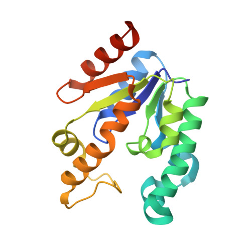
4BQS

Crystal structure of Mycobacterium tuberculosis shikimate kinase in complex with ADP and a shikimic acid derivative.


Experimental Data Snapshot

  • Method: X-RAY DIFFRACTION
  • Resolution: 2.15 Å
  • R-Value Free: 0.235 
  • R-Value Work: 0.172 
  • R-Value Observed: 0.176 

Starting Model: experimental
View more details

wwPDB Validation   3D Report Full Report


Ligand Structure Quality Assessment 


This is version 1.4 of the entry. See complete history


Literature

Mycobacterium tuberculosis shikimate kinase inhibitors: design and simulation studies of the catalytic turnover.

Blanco, B.Prado, V.Lence, E.Otero, J.M.Garcia-Doval, C.van Raaij, M.J.Llamas-Saiz, A.L.Lamb, H.Hawkins, A.R.Gonzalez-Bello, C.

(2013) J Am Chem Soc 135: 12366-12376

  • DOI: https://doi.org/10.1021/ja405853p

  • PubMed Abstract: 

    Shikimate kinase (SK) is an essential enzyme in several pathogenic bacteria and does not have any counterpart in human cells, thus making it an attractive target for the development of new antibiotics. The key interactions of the substrate and product binding and the enzyme movements that are essential for catalytic turnover of the Mycobacterium tuberculosis shikimate kinase enzyme (Mt-SK) have been investigated by structural and computational studies. Based on these studies several substrate analogs were designed and assayed. The crystal structure of Mt-SK in complex with ADP and one of the most potent inhibitors has been solved at 2.15 Å. These studies reveal that the fixation of the diaxial conformation of the C4 and C5 hydroxyl groups recognized by the enzyme or the replacement of the C3 hydroxyl group in the natural substrate by an amino group is a promising strategy for inhibition because it causes a dramatic reduction of the flexibility of the LID and shikimic acid binding domains. Molecular dynamics simulation studies showed that the product is expelled from the active site by three arginines (Arg117, Arg136, and Arg58). This finding represents a previously unknown key role of these conserved residues. These studies highlight the key role of the shikimic acid binding domain in the catalysis and provide guidance for future inhibitor designs.


  • Organizational Affiliation

    Centro Singular de Investigación en Química Biológica y Materiales Moleculares, Universidad de Santiago de Compostela, 15782 Santiago de Compostela, Spain.


Macromolecules
Find similar proteins by:  (by identity cutoff)  |  3D Structure
Entity ID: 1
MoleculeChains Sequence LengthOrganismDetailsImage
SHIKIMATE KINASE
A, B, C
176Mycobacterium tuberculosisMutation(s): 0 
EC: 2.7.1.71
UniProt
Find proteins for P9WPY3 (Mycobacterium tuberculosis (strain ATCC 25618 / H37Rv))
Explore P9WPY3 
Go to UniProtKB:  P9WPY3
Entity Groups  
Sequence Clusters30% Identity50% Identity70% Identity90% Identity95% Identity100% Identity
UniProt GroupP9WPY3
Sequence Annotations
Expand
  • Reference Sequence
Experimental Data & Validation

Experimental Data

  • Method: X-RAY DIFFRACTION
  • Resolution: 2.15 Å
  • R-Value Free: 0.235 
  • R-Value Work: 0.172 
  • R-Value Observed: 0.176 
  • Space Group: P 1 21 1
Unit Cell:
Length ( Å )Angle ( ˚ )
a = 40.653α = 90
b = 60.959β = 95.44
c = 89.976γ = 90
Software Package:
Software NamePurpose
REFMACrefinement
XDSdata reduction
SCALAdata scaling
MOLREPphasing

Structure Validation

View Full Validation Report


Ligand Structure Quality Assessment 


Entry History 

Deposition Data

Revision History  (Full details and data files)

  • Version 1.0: 2013-08-07
    Type: Initial release
  • Version 1.1: 2013-08-28
    Changes: Database references
  • Version 1.2: 2018-02-07
    Changes: Database references, Structure summary
  • Version 1.3: 2019-01-30
    Changes: Data collection, Experimental preparation
  • Version 1.4: 2023-12-20
    Changes: Data collection, Database references, Derived calculations, Other, Refinement description