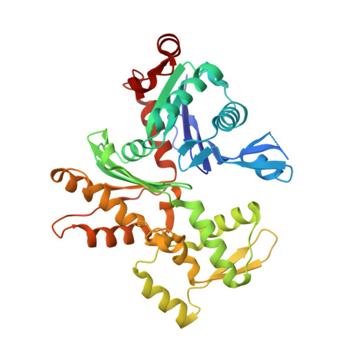
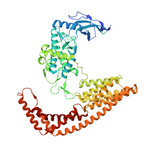
4CI6

Mechanisms of crippling actin-dependent phagocytosis by YopO


Experimental Data Snapshot

  • Method: X-RAY DIFFRACTION
  • Resolution: 2.65 Å
  • R-Value Free: 0.220 
  • R-Value Work: 0.186 
  • R-Value Observed: 0.187 

Starting Models: experimental
View more details

wwPDB Validation   3D Report Full Report


Ligand Structure Quality Assessment 


This is version 1.4 of the entry. See complete history


Literature

Yersinia Effector Yopo Uses Actin as Bait to Phosphorylate Proteins that Regulate Actin Polymerization.

Lee, W.L.Grimes, J.M.Robinson, R.C.

(2015) Nat Struct Mol Biol 22: 248

  • DOI: https://doi.org/10.1038/nsmb.2964
  • Primary Citation of Related Structures:  
    4CI6

  • PubMed Abstract: 

    Pathogenic Yersinia species evade host immune systems through the injection of Yersinia outer proteins (Yops) into phagocytic cells. One Yop, YopO, also known as YpkA, induces actin-filament disruption, impairing phagocytosis. Here we describe the X-ray structure of Yersinia enterocolitica YopO in complex with actin, which reveals that YopO binds to an actin monomer in a manner that blocks polymerization yet allows the bound actin to interact with host actin-regulating proteins. SILAC-MS and biochemical analyses confirm that actin-polymerization regulators such as VASP, EVL, WASP, gelsolin and the formin diaphanous 1 are directly sequestered and phosphorylated by YopO through formation of ternary complexes with actin. This leads to a model in which YopO at the membrane sequesters actin from polymerization while using the bound actin as bait to recruit, phosphorylate and misregulate host actin-regulating proteins to disrupt phagocytosis.


  • Organizational Affiliation

    1] Institute of Molecular and Cell Biology, Agency for Science, Technology and Research (A*STAR), Singapore. [2] Division of Structural Biology, Wellcome Trust Centre for Human Genetics, University of Oxford, Oxford, UK.


Macromolecules
Find similar proteins by:  (by identity cutoff)  |  3D Structure
Entity ID: 1
MoleculeChains Sequence LengthOrganismDetailsImage
ACTIN376Spodoptera frugiperdaMutation(s): 0 
UniProt
Find proteins for G3CKA6 (Spodoptera frugiperda)
Explore G3CKA6 
Go to UniProtKB:  G3CKA6
Entity Groups  
Sequence Clusters30% Identity50% Identity70% Identity90% Identity95% Identity100% Identity
UniProt GroupG3CKA6
Sequence Annotations
Expand
  • Reference Sequence
Find similar proteins by:  (by identity cutoff)  |  3D Structure
Entity ID: 2
MoleculeChains Sequence LengthOrganismDetailsImage
PROTEIN KINASE YOPO643Yersinia enterocoliticaMutation(s): 5 
EC: 2.7.11.1
UniProt
Find proteins for Q93KQ6 (Yersinia enterocolitica)
Explore Q93KQ6 
Go to UniProtKB:  Q93KQ6
Entity Groups  
Sequence Clusters30% Identity50% Identity70% Identity90% Identity95% Identity100% Identity
UniProt GroupQ93KQ6
Sequence Annotations
Expand
  • Reference Sequence
Experimental Data & Validation

Experimental Data

  • Method: X-RAY DIFFRACTION
  • Resolution: 2.65 Å
  • R-Value Free: 0.220 
  • R-Value Work: 0.186 
  • R-Value Observed: 0.187 
  • Space Group: P 1 21 1
Unit Cell:
Length ( Å )Angle ( ˚ )
a = 70.799α = 90
b = 121.85β = 94.87
c = 89.339γ = 90
Software Package:
Software NamePurpose
PHENIXrefinement
HKL-2000data reduction
SCALEPACKdata scaling
PHASERphasing

Structure Validation

View Full Validation Report


Ligand Structure Quality Assessment 


Entry History 

Deposition Data

Revision History  (Full details and data files)

  • Version 1.0: 2015-01-28
    Type: Initial release
  • Version 1.1: 2015-02-11
    Changes: Database references
  • Version 1.2: 2015-03-04
    Changes: Database references
  • Version 1.3: 2015-03-18
    Changes: Database references
  • Version 1.4: 2023-12-20
    Changes: Data collection, Database references, Derived calculations, Other, Refinement description