4D5J

Hypocrea jecorina cellobiohydrolase Cel7A E217Q soaked with xylotriose.


Experimental Data Snapshot

  • Method: X-RAY DIFFRACTION
  • Resolution: 1.50 Å
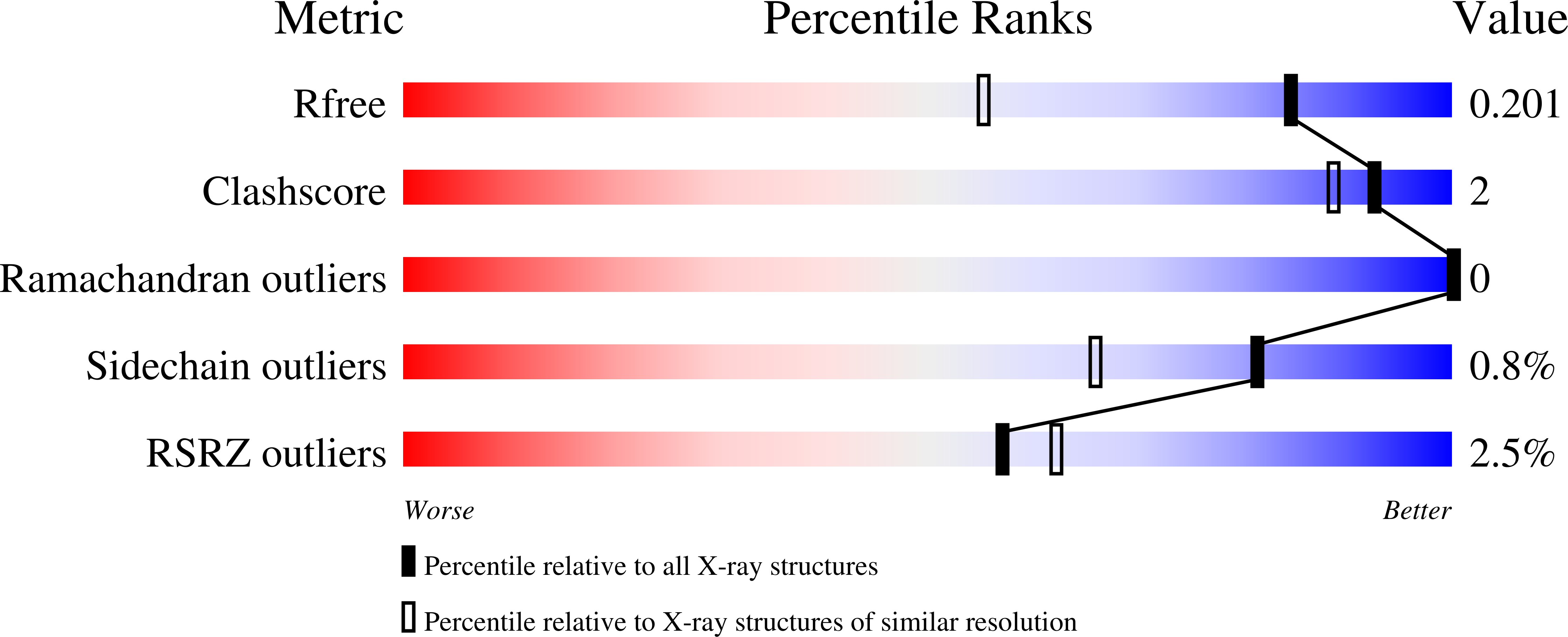
  • R-Value Free: 0.199 
  • R-Value Work: 0.171 
  • R-Value Observed: 0.173 

Starting Model: experimental
View more details

wwPDB Validation   3D Report Full Report


Ligand Structure Quality Assessment 


This is version 3.2 of the entry. See complete history


Literature

Structural Insights Into the Inhibition of Cellobiohydrolase Cel7A by Xylooligosaccharides.

Haddad Momeni, M.Ubhayasekera, W.Sandgren, M.Stahlberg, J.Hansson, H.

(2015) FEBS J 282: 2167

  • DOI: https://doi.org/10.1111/febs.13265
  • Primary Citation of Related Structures:  
    4D5I, 4D5J, 4D5O, 4D5P, 4D5Q, 4D5V

  • PubMed Abstract: 

    The filamentous fungus Hypocrea jecorina (anamorph of Trichoderma reesei) is the predominant source of enzymes for industrial saccharification of lignocellulose biomass. The major enzyme, cellobiohydrolase Cel7A, constitutes nearly half of the total protein in the secretome. The performance of such enzymes is susceptible to inhibition by compounds liberated by physico-chemical pre-treatment if the biomass is kept unwashed. Xylan and xylo-oligosaccharides (XOS) have been proposed to play a key role in inhibition of cellobiohydrolases of glycoside hydrolase family 7. To elucidate the mechanism behind this inhibition at a molecular level, we used X-ray crystallography to determine structures of H. jecorina Cel7A in complex with XOS. Structures with xylotriose, xylotetraose and xylopentaose revealed a predominant binding mode at the entrance of the substrate-binding tunnel of the enzyme, in which each xylose residue is shifted ~ 2.4 Å towards the catalytic center compared with binding of cello-oligosaccharides. Furthermore, partial occupancy of two consecutive xylose residues at subsites -2 and -1 suggests an alternative binding mode for XOS in the vicinity of the catalytic center. Interestingly, the -1 xylosyl unit exhibits an open aldehyde conformation in one of the structures and a ring-closed pyranoside in another complex. Complementary inhibition studies with p-nitrophenyl lactoside as substrate indicate mixed inhibition rather than pure competitive inhibition. The atomic coordinates and structure factors are available in the Protein Data Bank under accession number 4D5I (H. jecorina Cel7A E212Q variant, complex with xylotriose), 4D5J (H. jecorina Cel7A E217Q variant, complex with xylotriose), 4D5O (H. jecorina Cel7A E212Q variant, complex with xylopentaose), 4D5P (H. jecorina Cel7A E217Q variant, complex with xylopentaose), 4D5Q (wild-type H. jecorina Cel7A, complex with xylopentaose) and 4D5V (H. jecorina Cel7A E217Q variant, complex with xylotetraose).


  • Organizational Affiliation

    Department of Chemistry and Biotechnology, Swedish University of Agricultural Sciences, Uppsala, Sweden.


Macromolecules
Find similar proteins by:  (by identity cutoff)  |  3D Structure
Entity ID: 1
MoleculeChains Sequence LengthOrganismDetailsImage
CELLULOSE 1,4-BETA-CELLOBIOSIDASE434Trichoderma reeseiMutation(s): 1 
EC: 3.2.1.176 (PDB Primary Data), 3.2.1.91 (UniProt)
UniProt
Find proteins for P62694 (Hypocrea jecorina)
Explore P62694 
Go to UniProtKB:  P62694
Entity Groups  
Sequence Clusters30% Identity50% Identity70% Identity90% Identity95% Identity100% Identity
UniProt GroupP62694
Glycosylation
Glycosylation Sites: 1
Sequence Annotations
Expand
  • Reference Sequence
Oligosaccharides

Help

Entity ID: 2
MoleculeChains Length2D Diagram Glycosylation3D Interactions
beta-D-xylopyranose-(1-4)-beta-D-xylopyranose-(1-4)-beta-D-xylopyranose
B
3N/A
Glycosylation Resources
GlyTouCan:  G87429QT
GlyCosmos:  G87429QT
Small Molecules
Biologically Interesting Molecules (External Reference) 1 Unique
Experimental Data & Validation

Experimental Data

  • Method: X-RAY DIFFRACTION
  • Resolution: 1.50 Å
  • R-Value Free: 0.199 
  • R-Value Work: 0.171 
  • R-Value Observed: 0.173 
  • Space Group: I 2 2 2
Unit Cell:
Length ( Å )Angle ( ˚ )
a = 83.232α = 90
b = 83.083β = 90
c = 110.66γ = 90
Software Package:
Software NamePurpose
REFMACrefinement
XDSdata reduction
SCALAdata scaling
REFMACphasing

Structure Validation

View Full Validation Report


Ligand Structure Quality Assessment 


Entry History 

Deposition Data

Revision History  (Full details and data files)

  • Version 1.0: 2015-03-25
    Type: Initial release
  • Version 1.1: 2015-06-17
    Changes: Database references
  • Version 1.2: 2018-01-17
    Changes: Data collection
  • Version 1.3: 2019-05-08
    Changes: Data collection, Derived calculations, Experimental preparation
  • Version 2.0: 2020-03-11
    Changes: Data collection, Other, Polymer sequence
  • Version 3.0: 2020-07-29
    Type: Remediation
    Reason: Carbohydrate remediation
    Changes: Atomic model, Data collection, Derived calculations, Structure summary
  • Version 3.1: 2023-12-20
    Changes: Data collection, Database references, Derived calculations, Refinement description, Structure summary
  • Version 3.2: 2024-11-13
    Changes: Structure summary