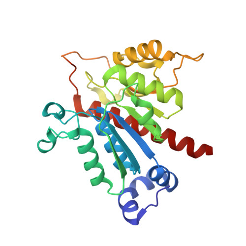
4D7A

Crystal structure of E. coli tRNA N6-threonylcarbamoyladenosine dehydratase, TcdA, in complex with AMP at 1.801 Angstroem resolution


Experimental Data Snapshot

  • Method: X-RAY DIFFRACTION
  • Resolution: 1.80 Å
  • R-Value Free: 0.177 
  • R-Value Work: 0.139 
  • R-Value Observed: 0.141 

Starting Model: experimental
View more details

wwPDB Validation   3D Report Full Report


Ligand Structure Quality Assessment 


This is version 1.1 of the entry. See complete history


Literature

The Crystal Structure and Small-Angle X-Ray Analysis of Csdl/Tcda Reveal a New tRNA Binding Motif in the Moeb/E1 Superfamily.

Lopez-Estepa, M.Arda, A.Savko, M.Round, A.Shepard, W.E.Bruix, M.Coll, M.Fernandez, F.J.Jimenez-Barbero, J.Vega, M.C.

(2015) PLoS One 10: 18606

  • DOI: https://doi.org/10.1371/journal.pone.0118606
  • Primary Citation of Related Structures:  
    4D79, 4D7A

  • PubMed Abstract: 

    Cyclic N6-threonylcarbamoyladenosine ('cyclic t6A', ct(6)A) is a non-thiolated hypermodification found in transfer RNAs (tRNAs) in bacteria, protists, fungi and plants. In bacteria and yeast cells ct(6)A has been shown to enhance translation fidelity and efficiency of ANN codons by improving the faithful discrimination of aminoacylated tRNAs by the ribosome. To further the understanding of ct(6)A biology we have determined the high-resolution crystal structures of CsdL/TcdA in complex with AMP and ATP, an E1-like activating enzyme from Escherichia coli, which catalyzes the ATP-dependent dehydration of t6A to form ct(6)A. CsdL/TcdA is a dimer whose structural integrity and dimer interface depend critically on strongly bound K+ and Na+ cations. By using biochemical assays and small-angle X-ray scattering we show that CsdL/TcdA can associate with tRNA with a 1:1 stoichiometry and with the proper position and orientation for the cyclization of t6A. Furthermore, we show by nuclear magnetic resonance that CsdL/TcdA engages in transient interactions with CsdA and CsdE, which, in the latter case, involve catalytically important residues. These short-lived interactions may underpin the precise channeling of sulfur atoms from cysteine to CsdL/TcdA as previously characterized. In summary, the combination of structural, biophysical and biochemical methods applied to CsdL/TcdA has afforded a more thorough understanding of how the structure of this E1-like enzyme has been fine tuned to accomplish ct(6)A synthesis on tRNAs while providing support for the notion that CsdA and CsdE are able to functionally interact with CsdL/TcdA.


  • Organizational Affiliation

    Chemical and Physical Biology, Center for Biological Research (CIB-CSIC), Madrid, Spain.


Macromolecules
Find similar proteins by:  (by identity cutoff)  |  3D Structure
Entity ID: 1
MoleculeChains Sequence LengthOrganismDetailsImage
TRNA THREONYLCARBAMOYLADENOSINE DEHYDRATASE
A, B, C, D
268Escherichia coli K-12Mutation(s): 0 
EC: 6.1
UniProt
Find proteins for Q46927 (Escherichia coli (strain K12))
Explore Q46927 
Go to UniProtKB:  Q46927
Entity Groups  
Sequence Clusters30% Identity50% Identity70% Identity90% Identity95% Identity100% Identity
UniProt GroupQ46927
Sequence Annotations
Expand
  • Reference Sequence
Small Molecules
Ligands 5 Unique
IDChains Name / Formula / InChI Key2D Diagram3D Interactions
AMP
Query on AMP

Download Ideal Coordinates CCD File 
F [auth A],
K [auth B],
Q [auth D]
ADENOSINE MONOPHOSPHATE
C10 H14 N5 O7 P
UDMBCSSLTHHNCD-KQYNXXCUSA-N
PO4
Query on PO4

Download Ideal Coordinates CCD File 
O [auth C]PHOSPHATE ION
O4 P
NBIIXXVUZAFLBC-UHFFFAOYSA-K
GOL
Query on GOL

Download Ideal Coordinates CCD File 
G [auth A]
H [auth A]
L [auth B]
R [auth D]
S [auth D]
G [auth A],
H [auth A],
L [auth B],
R [auth D],
S [auth D],
T [auth D]
GLYCEROL
C3 H8 O3
PEDCQBHIVMGVHV-UHFFFAOYSA-N
K
Query on K

Download Ideal Coordinates CCD File 
E [auth A],
I [auth B],
M [auth C],
P [auth D]
POTASSIUM ION
K
NPYPAHLBTDXSSS-UHFFFAOYSA-N
NA
Query on NA

Download Ideal Coordinates CCD File 
J [auth B],
N [auth C]
SODIUM ION
Na
FKNQFGJONOIPTF-UHFFFAOYSA-N
Experimental Data & Validation

Experimental Data

  • Method: X-RAY DIFFRACTION
  • Resolution: 1.80 Å
  • R-Value Free: 0.177 
  • R-Value Work: 0.139 
  • R-Value Observed: 0.141 
  • Space Group: P 1 21 1
Unit Cell:
Length ( Å )Angle ( ˚ )
a = 65.69α = 90
b = 97.16β = 111.61
c = 83.19γ = 90
Software Package:
Software NamePurpose
PHENIXrefinement
XDSdata reduction
Aimlessdata scaling

Structure Validation

View Full Validation Report


Ligand Structure Quality Assessment 


Entry History 

Deposition Data

Revision History  (Full details and data files)

  • Version 1.0: 2015-05-06
    Type: Initial release
  • Version 1.1: 2023-12-20
    Changes: Data collection, Database references, Derived calculations, Other, Refinement description