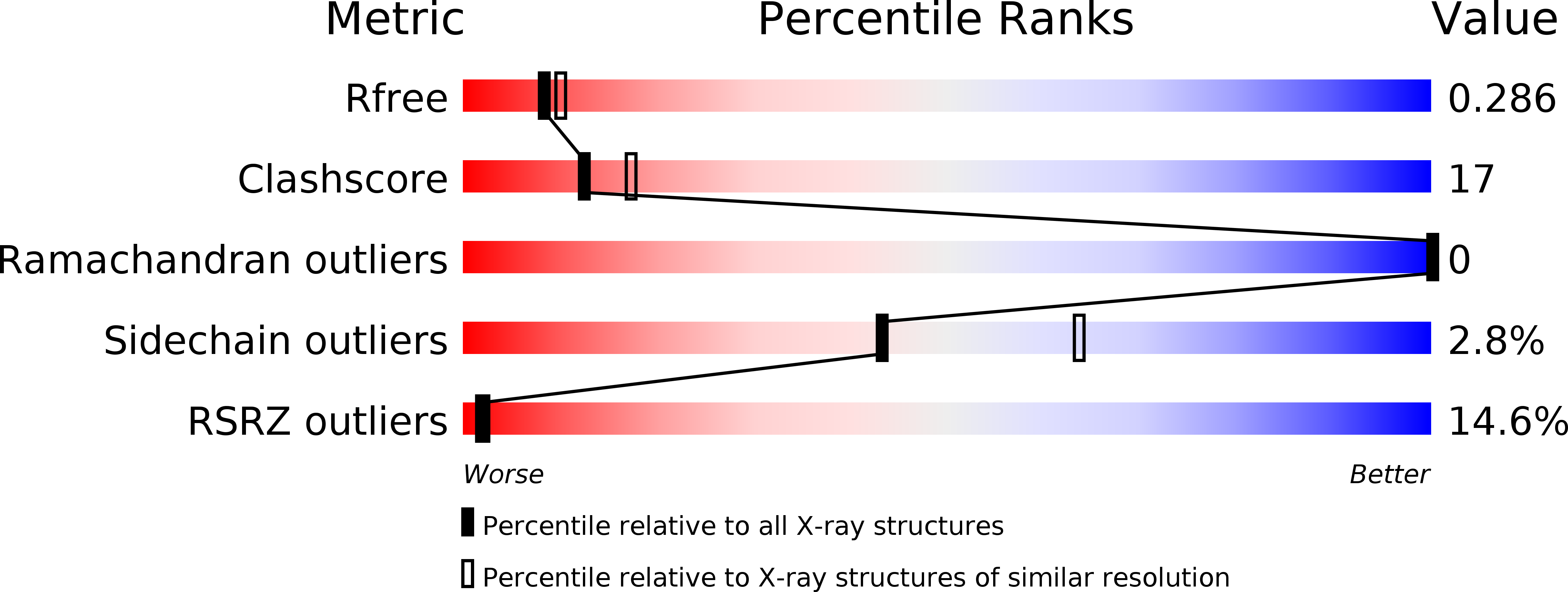
Structural studies on the folded domain of the human prion protein bound to the Fab fragment of the antibody POM1.
Baral, P.K., Wieland, B., Swayampakula, M., Polymenidou, M., Rahman, M.H., Kav, N.N., Aguzzi, A., James, M.N.(2012) Acta Crystallogr D Biol Crystallogr 68: 1501-1512
- PubMed: 23090399
- DOI: https://doi.org/10.1107/S0907444912037328
- Primary Citation of Related Structures:
4DGI - PubMed Abstract:
Prion diseases are neurodegenerative diseases characterized by the conversion of the cellular prion protein PrP(c) into a pathogenic isoform PrP(sc). Passive immunization with antiprion monoclonal antibodies can arrest the progression of prion diseases. Here, the crystal structure of the Fab fragment of an antiprion monoclonal antibody, POM1, in complex with human prion protein (huPrP(c)) has been determined to 2.4 Å resolution. The prion epitope of POM1 is in close proximity to the epitope recognized by the purportedly therapeutic antibody fragment ICSM18 Fab in complex with huPrP(c). POM1 Fab forms a 1:1 complex with huPrP(c) and the measured K(d) of 4.5 × 10(-7) M reveals moderately strong binding between them. Structural comparisons have been made among three prion-antibody complexes: POM1 Fab-huPrP(c), ICSM18 Fab-huPrP(c) and VRQ14 Fab-ovPrP(c). The prion epitopes recognized by ICSM18 Fab and VRQ14 Fab are adjacent to a prion glycosylation site, indicating possible steric hindrance and/or an altered binding mode to the glycosylated prion protein in vivo. However, both of the glycosylation sites on huPrP(c) are positioned away from the POM1 Fab binding epitope; thus, the binding mode observed in this crystal structure and the binding affinity measured for this antibody are most likely to be the same as those for the native prion protein in vivo.
Organizational Affiliation:
Department of Biochemistry, School of Translational Medicine, Faculty of Medicine and Dentistry, University of Alberta, Edmonton AB T6G 2H7, Canada.


