4ECM

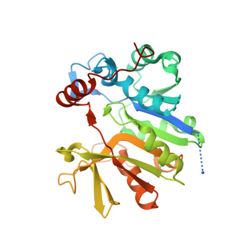
2.3 Angstrom Crystal Structure of a Glucose-1-phosphate Thymidylyltransferase from Bacillus anthracis in Complex with Thymidine-5-diphospho-alpha-D-glucose and Pyrophosphate


Experimental Data Snapshot

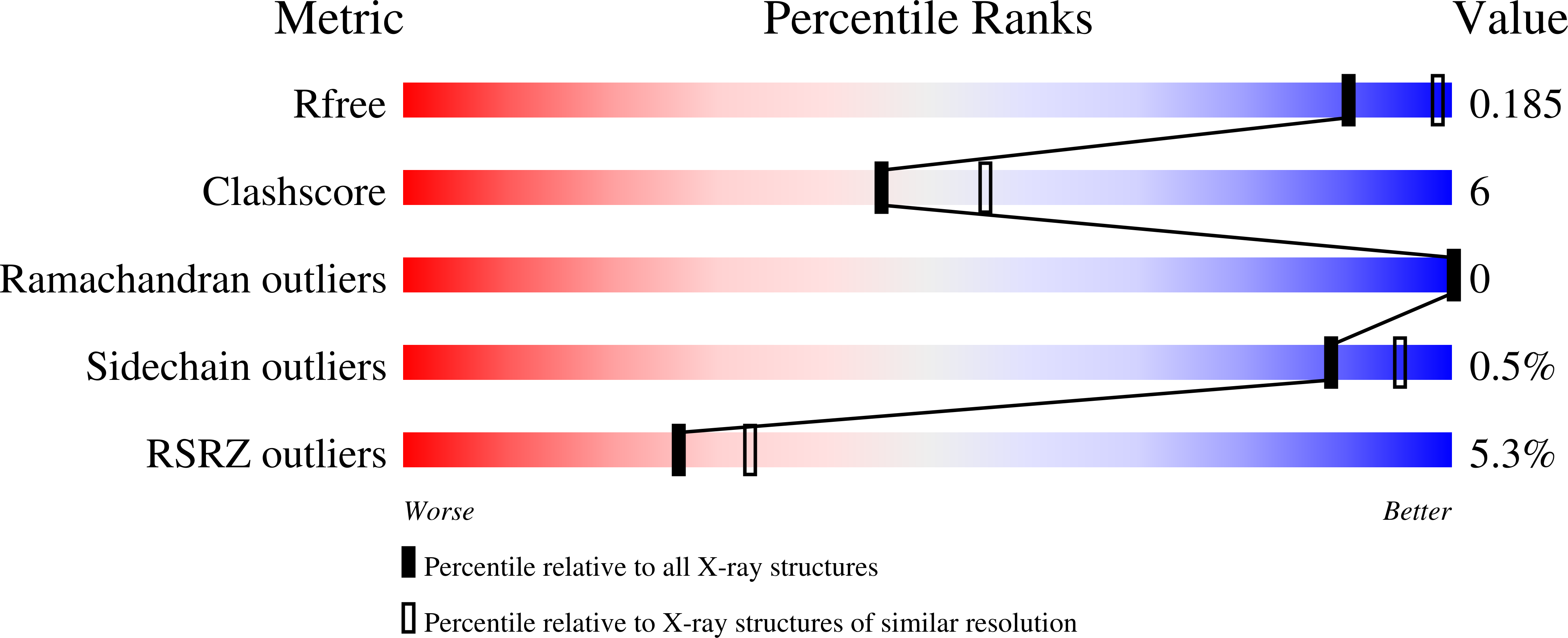
  • Method: X-RAY DIFFRACTION
  • Resolution: 2.30 Å
  • R-Value Free: 0.186 
  • R-Value Work: 0.153 
  • R-Value Observed: 0.154 

Starting Model: experimental
View more details

wwPDB Validation   3D Report Full Report


Ligand Structure Quality Assessment 


This is version 1.3 of the entry. See complete history


Literature

Structure of the Bacillus anthracis dTDP-L-rhamnose-biosynthetic enzyme glucose-1-phosphate thymidylyltransferase (RfbA).

Baumgartner, J.Lee, J.Halavaty, A.S.Minasov, G.Anderson, W.F.Kuhn, M.L.

(2017) Acta Crystallogr F Struct Biol Commun 73: 621-628

  • DOI: https://doi.org/10.1107/S2053230X17015357
  • Primary Citation of Related Structures:  
    4ECM

  • PubMed Abstract: 

    L-Rhamnose is a ubiquitous bacterial cell-wall component. The biosynthetic pathway for its precursor dTDP-L-rhamnose is not present in humans, which makes the enzymes of the pathway potential drug targets. In this study, the three-dimensional structure of the first protein of this pathway, glucose-1-phosphate thymidylyltransferase (RfbA), from Bacillus anthracis was determined. In other organisms this enzyme is referred to as RmlA. RfbA was co-crystallized with the products of the enzymatic reaction, dTDP-α-D-glucose and pyrophosphate, and its structure was determined at 2.3 Å resolution. This is the first reported thymidylyltransferase structure from a Gram-positive bacterium. RfbA shares overall structural characteristics with known RmlA homologs. However, RfbA exhibits a shorter sequence at its C-terminus, which results in the absence of three α-helices involved in allosteric site formation. Consequently, RfbA was observed to exhibit a quaternary structure that is unique among currently reported glucose-1-phosphate thymidylyltransferase bacterial homologs. These structural analyses suggest that RfbA may not be allosterically regulated in some organisms and is structurally distinct from other RmlA homologs.


  • Organizational Affiliation

    Department of Chemistry and Biochemistry, San Francisco State University, USA.


Macromolecules
Find similar proteins by:  (by identity cutoff)  |  3D Structure
Entity ID: 1
MoleculeChains Sequence LengthOrganismDetailsImage
Glucose-1-phosphate thymidylyltransferase269Bacillus anthracisMutation(s): 0 
Gene Names: BA1228BAS1135BA_1228GBAA_1228
EC: 2.7.7.24
UniProt
Find proteins for A0A6L8PCC3 (Bacillus anthracis)
Explore A0A6L8PCC3 
Go to UniProtKB:  A0A6L8PCC3
Entity Groups  
Sequence Clusters30% Identity50% Identity70% Identity90% Identity95% Identity100% Identity
UniProt GroupA0A6L8PCC3
Sequence Annotations
Expand
  • Reference Sequence
Small Molecules
Experimental Data & Validation

Experimental Data

Unit Cell:
Length ( Å )Angle ( ˚ )
a = 135.072α = 90
b = 135.072β = 90
c = 85.138γ = 120
Software Package:
Software NamePurpose
Blu-Icedata collection
PHASERphasing
REFMACrefinement
HKL-3000data reduction
HKL-3000data scaling

Structure Validation

View Full Validation Report


Ligand Structure Quality Assessment 


Entry History 

Revision History  (Full details and data files)

  • Version 1.0: 2012-04-04
    Type: Initial release
  • Version 1.1: 2012-05-23
    Changes: Atomic model
  • Version 1.2: 2017-11-15
    Changes: Database references, Refinement description
  • Version 1.3: 2023-09-13
    Changes: Data collection, Database references, Derived calculations, Refinement description