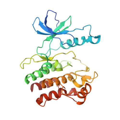
4F0F

Crystal Structure of the Roco4 Kinase Domain bound to AppCp from D. discoideum


Experimental Data Snapshot

  • Method: X-RAY DIFFRACTION
  • Resolution: 1.80 Å
  • R-Value Free: 0.245 
  • R-Value Work: 0.195 
  • R-Value Observed: 0.198 

Starting Model: experimental
View more details

wwPDB Validation   3D Report Full Report


Ligand Structure Quality Assessment 


This is version 1.2 of the entry. See complete history


Literature

Roco kinase structures give insights into the mechanism of Parkinson disease-related leucine-rich-repeat kinase 2 mutations.

Gilsbach, B.K.Ho, F.Y.Vetter, I.R.van Haastert, P.J.Wittinghofer, A.Kortholt, A.

(2012) Proc Natl Acad Sci U S A 109: 10322-10327

  • DOI: https://doi.org/10.1073/pnas.1203223109
  • Primary Citation of Related Structures:  
    4F0F, 4F0G, 4F1M, 4F1O, 4F1T

  • PubMed Abstract: 

    Mutations in human leucine-rich-repeat kinase 2 (LRRK2) have been found to be the most frequent cause of late-onset Parkinson disease. Here we show that Dictyostelium discoideum Roco4 is a suitable model to study the structural and biochemical characteristics of the LRRK2 kinase and can be used for optimization of current and identification of new LRRK2 inhibitors. We have solved the structure of Roco4 kinase wild-type, Parkinson disease-related mutants G1179S and L1180T (G2019S and I2020T in LRRK2) and the structure of Roco4 kinase in complex with the LRRK2 inhibitor H1152. Taken together, our data give important insight in the LRRK2 activation mechanism and, most importantly, explain the G2019S-related increase in LRRK2 kinase activity.


  • Organizational Affiliation

    Department of Cell Biochemistry, University of Groningen, 9747 AG, Groningen, The Netherlands.


Macromolecules
Find similar proteins by:  (by identity cutoff)  |  3D Structure
Entity ID: 1
MoleculeChains Sequence LengthOrganismDetailsImage
Serine/threonine-protein kinase roco4287Dictyostelium discoideumMutation(s): 0 
Gene Names: DDB_G0288251roco4
EC: 2.7.11.1
UniProt
Find proteins for Q6XHB2 (Dictyostelium discoideum)
Explore Q6XHB2 
Go to UniProtKB:  Q6XHB2
Entity Groups  
Sequence Clusters30% Identity50% Identity70% Identity90% Identity95% Identity100% Identity
UniProt GroupQ6XHB2
Sequence Annotations
Expand
  • Reference Sequence
Small Molecules
Ligands 1 Unique
IDChains Name / Formula / InChI Key2D Diagram3D Interactions
ACP
Query on ACP

Download Ideal Coordinates CCD File 
B [auth A]PHOSPHOMETHYLPHOSPHONIC ACID ADENYLATE ESTER
C11 H18 N5 O12 P3
UFZTZBNSLXELAL-IOSLPCCCSA-N
Experimental Data & Validation

Experimental Data

  • Method: X-RAY DIFFRACTION
  • Resolution: 1.80 Å
  • R-Value Free: 0.245 
  • R-Value Work: 0.195 
  • R-Value Observed: 0.198 
  • Space Group: P 43 21 2
Unit Cell:
Length ( Å )Angle ( ˚ )
a = 42.72α = 90
b = 42.72β = 90
c = 339.46γ = 90
Software Package:
Software NamePurpose
PROdata collection
PHASERphasing
REFMACrefinement
XDSdata reduction
XSCALEdata scaling

Structure Validation

View Full Validation Report


Ligand Structure Quality Assessment 


Entry History 

Deposition Data

Revision History  (Full details and data files)

  • Version 1.0: 2012-06-27
    Type: Initial release
  • Version 1.1: 2012-07-11
    Changes: Database references
  • Version 1.2: 2023-09-13
    Changes: Data collection, Database references, Derived calculations, Refinement description