4GJS

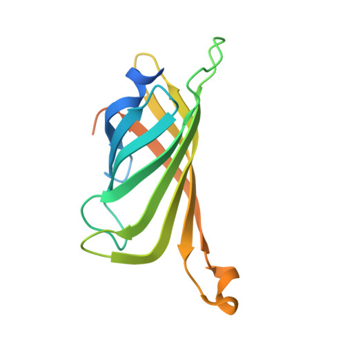
Streptavidin-K121H


Experimental Data Snapshot

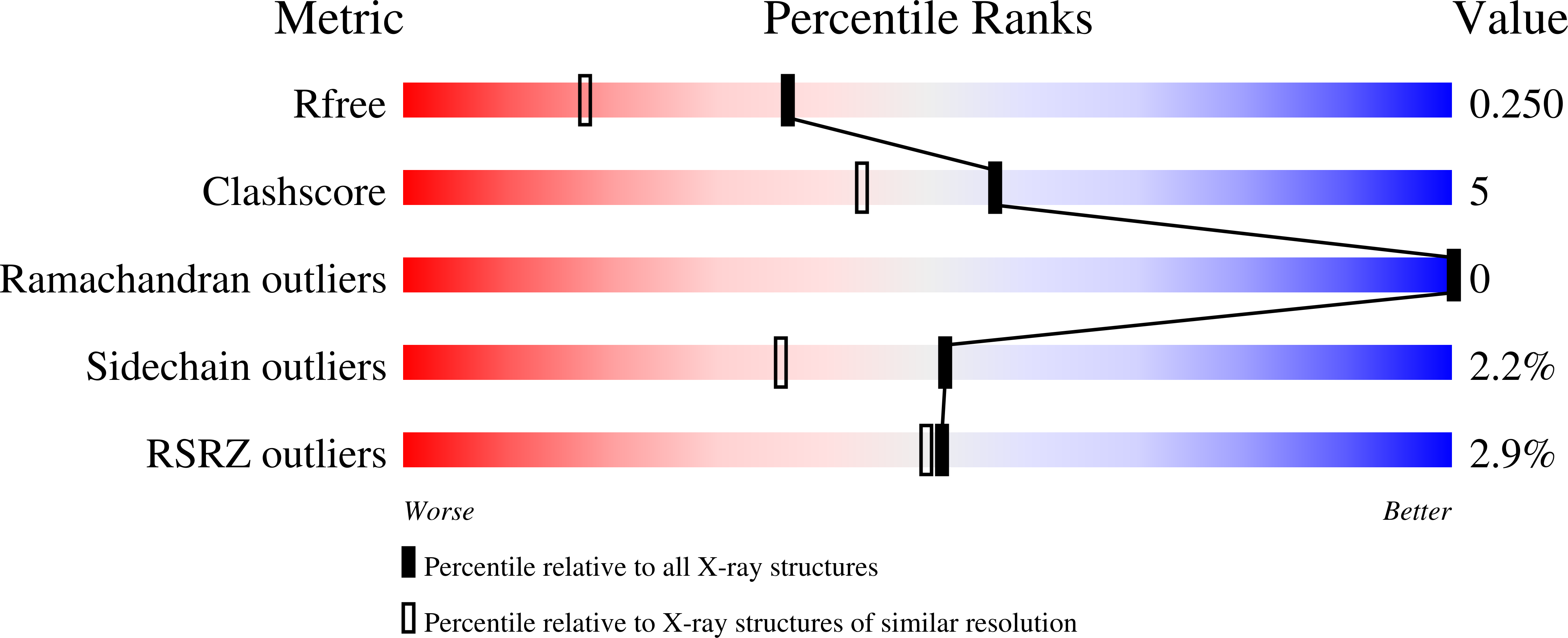
  • Method: X-RAY DIFFRACTION
  • Resolution: 1.85 Å
  • R-Value Free: 0.250 
  • R-Value Work: 0.203 
  • R-Value Observed: 0.205 

wwPDB Validation   3D Report Full Report


Ligand Structure Quality Assessment 


This is version 1.3 of the entry. See complete history


Literature

A dual anchoring strategy for the localization and activation of artificial metalloenzymes based on the biotin-streptavidin technology.

Zimbron, J.M.Heinisch, T.Schmid, M.Hamels, D.Nogueira, E.S.Schirmer, T.Ward, T.R.

(2013) J Am Chem Soc 135: 5384-5388

  • DOI: https://doi.org/10.1021/ja309974s
  • Primary Citation of Related Structures:  
    4GJS, 4GJV

  • PubMed Abstract: 

    Artificial metalloenzymes result from anchoring an active catalyst within a protein environment. Toward this goal, various localization strategies have been pursued: covalent, supramolecular, or dative anchoring. Herein we show that introduction of a suitably positioned histidine residue contributes to firmly anchor, via a dative bond, a biotinylated rhodium piano stool complex within streptavidin. The in silico design of the artificial metalloenzyme was confirmed by X-ray crystallography. The resulting artificial metalloenzyme displays significantly improved catalytic performance, both in terms of activity and selectivity in the transfer hydrogenation of imines. Depending on the position of the histidine residue, both enantiomers of the salsolidine product can be obtained.


  • Organizational Affiliation

    Biozentrum and Department of Chemistry, University of Basel, CH-4056 Basel, Switzerland.


Macromolecules
Find similar proteins by:  (by identity cutoff)  |  3D Structure
Entity ID: 1
MoleculeChains Sequence LengthOrganismDetailsImage
Streptavidin
A, B
159Streptomyces avidiniiMutation(s): 1 
UniProt
Find proteins for P22629 (Streptomyces avidinii)
Explore P22629 
Go to UniProtKB:  P22629
Entity Groups  
Sequence Clusters30% Identity50% Identity70% Identity90% Identity95% Identity100% Identity
UniProt GroupP22629
Sequence Annotations
Expand
  • Reference Sequence
Experimental Data & Validation

Experimental Data

  • Method: X-RAY DIFFRACTION
  • Resolution: 1.85 Å
  • R-Value Free: 0.250 
  • R-Value Work: 0.203 
  • R-Value Observed: 0.205 
  • Space Group: C 1 2 1
Unit Cell:
Length ( Å )Angle ( ˚ )
a = 81.652α = 90
b = 81.331β = 104.83
c = 47.002γ = 90
Software Package:
Software NamePurpose
SCALAdata scaling
PHENIXrefinement

Structure Validation

View Full Validation Report


Ligand Structure Quality Assessment 


Entry History 

Deposition Data

Revision History  (Full details and data files)

  • Version 1.0: 2013-02-13
    Type: Initial release
  • Version 1.1: 2013-04-10
    Changes: Database references
  • Version 1.2: 2013-04-24
    Changes: Database references
  • Version 1.3: 2024-02-28
    Changes: Data collection, Database references, Derived calculations, Refinement description