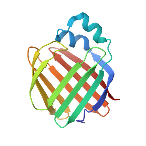
4GKC

Crystal structure of Q108K:K40L:T51V:T53C:R58W:T29L:Y19W:Q4A mutant of cellular retinol binding protein II complex with all-trans-retinal at 1.33


Experimental Data Snapshot

  • Method: X-RAY DIFFRACTION
  • Resolution: 1.30 Å
  • R-Value Free: 0.225 
  • R-Value Work: 0.187 
  • R-Value Observed: 0.189 

Starting Model: experimental
View more details

wwPDB Validation   3D Report Full Report


Ligand Structure Quality Assessment 


This is version 1.3 of the entry. See complete history


Literature

Tuning the electronic absorption of protein-embedded all-trans-retinal.

Wang, W.Nossoni, Z.Berbasova, T.Watson, C.T.Yapici, I.Lee, K.S.Vasileiou, C.Geiger, J.H.Borhan, B.

(2012) Science 338: 1340-1343

  • DOI: https://doi.org/10.1126/science.1226135
  • Primary Citation of Related Structures:  
    4EDE, 4EEJ, 4EFG, 4EXZ, 4GKC, 4RUU

  • PubMed Abstract: 

    Protein-chromophore interactions are a central component of a wide variety of critical biological processes such as color vision and photosynthesis. To understand the fundamental elements that contribute to spectral tuning of a chromophore inside the protein cavity, we redesigned human cellular retinol binding protein II (hCRBPII) to fully encapsulate all-trans-retinal and form a covalent bond as a protonated Schiff base. This system, using rational mutagenesis designed to alter the electrostatic environment within the binding pocket of the host protein, enabled regulation of the absorption maximum of the pigment in the range of 425 to 644 nanometers. With only nine point mutations, the hCRBPII mutants induced a systematic shift in the absorption profile of all-trans-retinal of more than 200 nanometers across the visible spectrum.


  • Organizational Affiliation

    Department of Chemistry, Michigan State University, East Lansing, MI 48824, USA.


Macromolecules
Find similar proteins by:  (by identity cutoff)  |  3D Structure
Entity ID: 1
MoleculeChains Sequence LengthOrganismDetailsImage
Retinol-binding protein 2
A, B
133Homo sapiensMutation(s): 8 
Gene Names: RBP2CRBP2
UniProt & NIH Common Fund Data Resources
Find proteins for P50120 (Homo sapiens)
Explore P50120 
Go to UniProtKB:  P50120
PHAROS:  P50120
GTEx:  ENSG00000114113 
Entity Groups  
Sequence Clusters30% Identity50% Identity70% Identity90% Identity95% Identity100% Identity
UniProt GroupP50120
Sequence Annotations
Expand
  • Reference Sequence
Small Molecules
Binding Affinity Annotations 
IDSourceBinding Affinity
RET PDBBind:  4GKC Kd: 150 (nM) from 1 assay(s)
Experimental Data & Validation

Experimental Data

  • Method: X-RAY DIFFRACTION
  • Resolution: 1.30 Å
  • R-Value Free: 0.225 
  • R-Value Work: 0.187 
  • R-Value Observed: 0.189 
  • Space Group: P 1
Unit Cell:
Length ( Å )Angle ( ˚ )
a = 30.97α = 86.1
b = 36.002β = 86.46
c = 64.016γ = 65.32
Software Package:
Software NamePurpose
HKL-2000data collection
MOLREPphasing
REFMACrefinement
HKL-2000data reduction
HKL-2000data scaling

Structure Validation

View Full Validation Report


Ligand Structure Quality Assessment 


Entry History 

Deposition Data

Revision History  (Full details and data files)

  • Version 1.0: 2012-12-26
    Type: Initial release
  • Version 1.1: 2018-01-24
    Changes: Advisory, Structure summary
  • Version 1.2: 2023-09-13
    Changes: Advisory, Data collection, Database references, Derived calculations, Refinement description
  • Version 1.3: 2024-11-20
    Changes: Structure summary